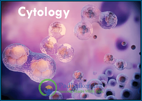
Introduction to Cytology
Cytology is the branch of biology that deals with the study of cells, their structure, function, and behavior. Cytologists study the various structures and functions of cells, including their organelles, membranes, and cytoplasm. They also investigate the ways in which cells interact with each other and with their environment.
Definition and Scope of Cytology
Cytology, derived from the Greek words “kytos” meaning “container” and “logos” meaning “study”, encompasses the investigation of cells in their various forms, structures, and functions. It is a discipline that delves deep into the microscopic intricacies of cells, offering an understanding of their diverse compositions and behaviors. Through cytology, we peer into the world of cells, unraveling their mysteries and unraveling the complexities that govern life’s processes.
The scope of cytology is vast and all-encompassing, transcending the boundaries of species and systems. It involves not only the observation of cell morphology but also the analysis of their functions, interactions, and deviations from the norm. Cytologists investigate the transformations cells undergo during development, growth, and response to external stimuli, making it a cornerstone of research in fields such as developmental biology, immunology, and oncology.
Importance of Cytology in Medical Diagnosis

Cytology occupies a critical position in the realm of medical diagnosis. By examining cells and their microstructures, cytology plays an indispensable role in identifying and characterizing diseases at the cellular level. Through techniques like exfoliative cytology and interventional cytology, medical professionals can glean invaluable information about the presence of abnormal cells, potential malignancies, and other health conditions.
Cytology is especially renowned for its contribution to cancer detection and management. By studying cellular changes indicative of malignancy, cytologists aid in the early diagnosis of cancers, enabling timely intervention and treatment. From Pap smears to fine needle aspiration, cytology has revolutionized cancer screening and monitoring, offering minimally invasive yet highly informative methods for clinicians and patients alike.
Types of Cytology
In the vast expanse of the microscopic universe, cytology offers a plethora of techniques to explore the intricate world of cells. From non-invasive procedures to comprehensive diagnostic interventions, cytology encompasses a range of methods that provide invaluable insights into cellular structures, behaviors, and anomalies.
Cytological techniques have evolved over the years, each designed to address specific questions related to cell structure, function, and health. These techniques enable researchers and medical practitioners to examine cells with varying degrees of detail, allowing for the identification of normal and abnormal cellular features. Here, we delve into two prominent categories of cytology techniques: exfoliative cytology and interventional cytology.
Exfoliative Cytology: Sampling and Analysis
Exfoliative cytology, often referred to as “cytology of the shed cells,” is a non-invasive method that involves collecting cells that have been naturally shed from the body’s surfaces. One of the most common applications of exfoliative cytology is the Pap smear, which plays a pivotal role in cervical cancer screening. By obtaining cervical cell samples and analyzing them under a microscope, cytologists can identify cellular changes that may indicate precancerous or cancerous conditions.
The simplicity and accessibility of exfoliative cytology make it a valuable tool in preventive healthcare. Beyond cervical cytology, exfoliative techniques are also used in diagnosing conditions like oral cancers and urinary tract infections. By analyzing cells obtained from these areas, healthcare professionals can promptly identify cellular abnormalities, enabling early intervention and treatment.
Interventional Cytology: Biopsy and Diagnostic Procedures
Interventional cytology, on the other hand, involves the extraction of cells from specific tissues or lesions for closer examination. This category includes procedures such as fine needle aspiration (FNA) and core needle biopsies. FNA, a minimally invasive technique, entails using a thin needle to extract cells from suspicious masses or tumors. The extracted cells are then analyzed to determine whether they exhibit features indicative of malignancy.
Core needle biopsies, while slightly more invasive, provide larger tissue samples for analysis. These biopsies are commonly employed when a deeper understanding of tissue architecture is required, aiding in the precise diagnosis of conditions like breast cancer and lymphomas. Interventional cytology’s ability to provide substantial tissue samples makes it an indispensable tool for confirming diagnoses and guiding treatment decisions.
Applications of Cytology
The world of cytology extends its reach far beyond the confines of the microscopic realm, with applications that have revolutionized disease detection and diagnosis. From unveiling the early stages of cancer to identifying a spectrum of medical conditions, cytology plays a pivotal role in providing valuable insights into the inner workings of cells. In this article, we explore the multifaceted applications of cytology, delving into its significant role in medical diagnosis.
Role of Cytology in Disease Detection
Cytology serves as a powerful tool in detecting diseases at their earliest stages, often before visible symptoms manifest. By analyzing cellular changes, cytologists can uncover potential health issues and alert healthcare professionals to the need for further investigation. This early intervention can significantly improve patient outcomes by enabling timely treatment and management.
Cytology Tests for Cancer Diagnosis

One of the most notable applications of cytology is in the realm of cancer diagnosis. Cytology tests are instrumental in identifying cancerous changes in cells, allowing for early detection and subsequent treatment. The Pap smear, for instance, has been a game-changer in cervical cancer screening. By identifying abnormal cellular features, such as precancerous lesions, healthcare providers can take preventive measures to halt the progression of the disease.
Furthermore, cytology is widely utilized in diagnosing cancers affecting various parts of the body. Fine needle aspiration (FNA) cytology allows for the non-invasive sampling of suspicious masses, providing crucial information about the nature of the tumor. This technique aids in determining whether the tumor is benign or malignant, guiding treatment decisions and interventions.
Other Medical Conditions Detected by Cytology
Beyond cancer, cytology’s diagnostic prowess extends to a range of medical conditions. In the field of hematology, for example, cytology plays a pivotal role in identifying blood disorders such as leukemia and lymphoma. By analyzing blood cells under a microscope, cytologists can identify abnormal cell populations and variations in cell morphology that indicate disease.
In addition, cytology is applied to the detection of infectious diseases. By examining cellular samples from affected areas, such as sputum samples from the respiratory tract, cytologists can identify the presence of pathogens and provide vital information for accurate diagnosis and treatment.
Performing Cytology Tests
Behind every cytology test lies a collaborative effort that involves skilled professionals, meticulous processes, and a commitment to accurate diagnosis. The journey from sample collection to diagnostic interpretation is a vital one, contributing to the detection and understanding of various health conditions. In this article, we explore the intricacies of performing cytology tests, shedding light on the roles of professionals, the testing process, and the art of cytologic evaluation.
Professionals Involved in Cytology Testing
Cytology testing is a multidisciplinary endeavor that involves a team of dedicated professionals, each contributing their expertise to different stages of the process. Cytotechnologists are specialized laboratory professionals who play a pivotal role in examining cellular samples under the microscope. Their trained eyes discern normal cellular patterns from abnormalities, aiding in the identification of diseases.
Pathologists, medical doctors with specialized training in diagnosing diseases, collaborate closely with cytotechnologists. They provide oversight, review complex cases, and make final diagnostic interpretations. Their expertise ensures accurate and clinically relevant diagnoses, guiding treatment decisions and patient management.
The Process of Conducting a Cytology Test
Cytology testing encompasses a series of steps designed to extract, process, and analyze cellular samples. The process begins with sample collection, which can range from a swab of a mucosal surface for exfoliative cytology to a biopsy for interventional cytology. Once collected, the samples undergo various processing steps to prepare them for examination under a microscope.
In the laboratory, cytotechnologists stain and prepare the samples on slides, making cellular features more visible for analysis. They meticulously study the cellular structures, noting any deviations from normal morphology. Pathologists review the cytotechnologist’s findings, corroborating interpretations and providing their expert opinion.
Cytologic Evaluation and Reporting
The heart of cytology lies in the evaluation and reporting of cellular findings. Cytotechnologists and pathologists evaluate cells based on predetermined criteria and classifications. Their combined assessments determine whether cells are normal, atypical, or indicative of disease. This evaluation serves as the basis for a detailed report that includes descriptions of cellular characteristics and any identified abnormalities.
Communication is key in the final phase of cytology testing. Pathologists carefully draft reports that not only describe cellular findings but also provide valuable clinical context. These reports are shared with referring physicians, forming a crucial link in the diagnostic chain. The information guides clinicians in making informed decisions about patient care and treatment strategies.
Cytology Test Procedure
In the realm of medical diagnostics, cytology tests stand as a powerful tool that offers a window into the microscopic world of cells. These tests provide crucial insights into various health conditions, aiding in early detection and accurate diagnosis. In this article, we delve into the intricate details of the cytology test procedure, from preparing for the test to the techniques used for collecting and analyzing cellular specimens.
Preparing for a Cytology Test

Before undergoing a cytology test, it’s important to be adequately prepared to ensure accurate results and a smooth testing process. Here are some key steps to consider:
Consultation with a Healthcare Provider: Discuss the reasons for the cytology test with your healthcare provider. Understand the purpose of the test, its potential outcomes, and its implications for your health.
Medication and Allergies: Inform your healthcare provider about any medications you are taking and any known allergies. Certain medications or conditions might affect the test results or the testing process itself.
Instructions for Sample Collection: Depending on the type of cytology test, you might receive specific instructions for sample collection. Follow these instructions carefully to ensure the validity of the results.
Clothing: Wear comfortable clothing that allows easy access to the area being tested, if applicable.
Discomfort or Anxiety: If you’re feeling anxious or have concerns about the procedure, communicate them to your healthcare provider. They can provide information and support to ease your worries.
The Fine Needle Aspiration Technique
Fine Needle Aspiration (FNA) is a widely used technique in cytology for obtaining cellular samples from lumps, masses, or suspicious lesions. This minimally invasive procedure involves the insertion of a thin needle into the targeted area to collect cellular material. FNA is often used to determine the nature of a lump, whether it is benign or potentially cancerous.
Here’s an overview of the FNA procedure:
Local Anesthesia: In some cases, a local anesthetic is administered to numb the area where the needle will be inserted. This helps minimize discomfort during the procedure.
Needle Insertion: A fine needle is inserted into the area of interest. The physician may use imaging techniques such as ultrasound or CT scans to guide the needle accurately.
Aspiration: The needle is gently moved back and forth within the mass to collect cellular material. Aspiration creates a vacuum that draws cells into the needle’s hollow core.
Sample Collection: The aspirated material is then placed on glass slides, smeared, and stained. The prepared slides are sent to the laboratory for cytological evaluation.
Collecting Cytological Specimens
Cytological specimens can be collected through various methods, depending on the area being tested and the type of cytology test. Some common methods include:
Swabs: Swabs are used to collect cellular material from mucosal surfaces, such as the cervix or oral cavity. The collected cells are then transferred onto slides for analysis.
Brushes: Brushes are employed to collect cells from internal surfaces, such as the bronchial passages. The brush is rotated to collect cells, which are then transferred onto slides.
Scrapings: Scrapings involve gently scraping the surface of a tissue or lesion to obtain cellular material. These scrapings are then processed and analyzed.
Biopsies: Biopsies involve the removal of a small piece of tissue for analysis. These tissue samples are processed and prepared for examination under a microscope.
Comparative Analysis
In the world of medical diagnostics, the quest for accurate and timely information is paramount. When it comes to investigating suspicious masses, lesions, or abnormalities, two primary diagnostic approaches come to the forefront: tissue biopsy and cytopathology. Each method offers distinct advantages and limitations, catering to various clinical scenarios and patient needs.
Differentiating Biopsy and Cytology Tests
Tissue Biopsy: A tissue biopsy involves the removal of a small piece of tissue from a suspicious area for detailed examination under a microscope. This approach provides a larger and more comprehensive sample of tissue compared to cytopathology. Tissue biopsies are often performed using techniques such as core needle biopsies or surgical excisions.
Cytopathology: It involves the examination of individual cells or small clusters of cells collected from a suspicious area. This method is non-invasive and often utilizes techniques like fine needle aspiration (FNA) or swabbing to obtain cellular samples.
Pros and Cons of Each Diagnostic Approach
Tissue Biopsy Pros:
Tissue Architecture: Tissue biopsies provide a more comprehensive view of cellular architecture, allowing pathologists to assess the organization and relationships between cells.
Tissue Characterization: Biopsies are particularly useful when detailed tissue characterization is required, such as differentiating between various types of tumors or assessing tissue integrity.
Molecular Analysis: Tissue biopsies offer the opportunity for molecular analysis, allowing for the detection of specific genetic mutations or markers.
Tissue Biopsy Cons:
Invasiveness: Tissue biopsies are more invasive compared to cytopathology techniques. The removal of tissue can lead to discomfort and potential complications.
Procedure Complexity: Some tissue biopsy procedures, especially surgical excisions, may require anesthesia and longer recovery times.
Sampling Limitation: Biopsy samples might not capture the full extent of heterogeneity in some tumors, potentially leading to sampling errors.
Cytopathology Pros:
Non-Invasive: Cytopathology techniques are minimally invasive and often do not require anesthesia. This makes them suitable for patients who might not tolerate more invasive procedures well.
Real-time Analysis: Cytopathology offers a quicker turnaround time for results compared to tissue biopsies, as cellular samples can be analyzed more rapidly.
Monitoring: Cytopathology can be used for regular monitoring of patients with known conditions, allowing for changes in cell characteristics to be tracked over time.
Cytopathology Cons:
Cellular Detail: While it offers insights into cellular characteristics, it might not provide the same level of tissue architecture information as tissue biopsies.
Limited Tissue: The amount of tissue obtained through cytopathology is limited, which can impact the ability to conduct certain tests or analyses.
Sampling Challenge: Sampling errors might occur due to the heterogeneity of lesions, as the selected sample might not accurately represent the entire lesion.
Advantages of Cytology
In the landscape of modern medical practice, the role of cytology stands as a beacon of innovation and diagnostic precision. Cytology, the study of cellular structures and functions, has garnered widespread recognition for its invaluable contributions to disease detection, diagnosis, and patient care. The benefits of Cytological Testing in Medical Practice are:
1. Early Disease Detection: One of the foremost advantages of cytology lies in its ability to detect diseases at their earliest stages. Cytological tests, such as Pap smears, can identify cellular abnormalities before visible symptoms manifest. This early detection empowers healthcare professionals to intervene promptly, leading to improved treatment outcomes and potentially preventing the progression of diseases like cancer.
2. Minimally Invasive: Cytological testing offers a non-invasive approach to obtaining diagnostic information. Techniques like fine needle aspiration (FNA) require only a thin needle to collect cellular samples, minimizing patient discomfort and reducing the risk of complications. This makes cytology a preferred choice for individuals who may be sensitive to invasive procedures.
3. Rapid Turnaround Time: In the fast-paced world of healthcare, timely results are of paramount importance. Cytology tests often offer a relatively quick turnaround time for results. This speed is especially valuable when immediate decisions about patient care are required, enabling healthcare providers to make informed choices without unnecessary delays.
4. Real-time Monitoring: Cytology’s non-invasive nature makes it conducive to repeated testing and monitoring of conditions over time. This is particularly advantageous for tracking disease progression, evaluating the effectiveness of treatments, and making necessary adjustments to patient care plans.
5. Cost-Efficiency: Cytology offers a cost-effective diagnostic approach when compared to more invasive procedures like biopsies. The reduced need for anesthesia, shorter recovery times, and the ability to perform tests on-site contribute to the cost-efficiency of cytological testing.
6. Accessibility and Reach: Cytology is accessible to a wide range of patients, regardless of age or health status. Screening programs utilizing cytological tests, such as cervical cancer screenings, have the potential to reach a large population and impact public health positively.
7. Diagnosing Infectious Diseases: Cytology has a role in diagnosing infectious diseases by identifying pathogens in cellular samples. By examining cells from areas such as respiratory tracts, cytologists can detect bacterial, viral, or fungal infections.
8. Precise Targeting: Cytological techniques like FNA allow for precise targeting of specific areas of concern, enabling healthcare providers to gather diagnostic information directly from the site of interest. This targeted approach minimizes the need for unnecessary sampling and reduces the risk of false negatives.
Essential Components for Effective Cytopathology Services
In the intricate world of medical diagnostics, cytopathology services stand as a cornerstone, providing critical insights into cellular structures and abnormalities. Behind the scenes of every successful cytopathology practice lies a foundation built upon essential components that ensure accuracy, reliability, and excellence in patient care.

Infrastructure:
1. Laboratory Facilities: An integral aspect of effective cytopathology services is a well-equipped laboratory. The laboratory should have state-of-the-art microscopes, staining equipment, and advanced imaging technology to facilitate accurate analysis of cellular samples. Adequate workspace and appropriate ventilation are also essential to ensure a safe and productive environment.
2. Skilled Workforce: A skilled and trained workforce is the backbone of any successful cytopathology service. This includes cytotechnologists who are trained to process and analyze cellular samples under the microscope, as well as pathologists with expertise in interpreting cytological findings. Collaboration between these professionals ensures accurate diagnoses.
3. Sample Handling and Processing: Efficient sample handling and processing are crucial to obtaining reliable results. Proper protocols for sample collection, fixation, staining, and slide preparation must be followed to maintain the integrity of cellular specimens and prevent artifacts.
4. Quality Control Measures: Implementing quality control measures is essential to ensure consistency and accuracy. Regular monitoring of equipment, staining techniques, and diagnostic processes helps identify and rectify any potential errors. External quality assurance programs and proficiency testing contribute to maintaining high standards of performance.
5. Information Management Systems: Effective information management systems are necessary to keep track of patient data, test results, and follow-up recommendations. Digital systems aid in accurate record-keeping, result reporting, and data analysis.
Quality Assurance and Training
1. Continuous Education: Regular training and continuing education programs are critical for maintaining a skilled workforce. Cytotechnologists and pathologists should stay updated on the latest advancements in cytopathology techniques, technologies, and diagnostic criteria.
2. Peer Review and Consultation: Instituting a system of peer review and consultation ensures a second opinion on challenging cases. Collaboration among pathologists fosters discussion and consensus on diagnoses, enhancing accuracy and diagnostic confidence.
3. Standard Operating Procedures: Establishing clear and standardized operating procedures helps maintain consistency in sample processing, analysis, and reporting. These procedures provide a framework for staff to follow, reducing the risk of errors.
4. Quality Audits: Regular quality audits evaluate the performance of the cytopathology service against established standards. These audits identify areas of improvement, measure compliance with protocols, and enhance overall quality.
5. Accreditation and Compliance: Seeking accreditation from relevant regulatory bodies, such as the College of American Pathologists (CAP), demonstrates a commitment to maintaining high-quality services. Compliance with accreditation standards ensures adherence to best practices.
Branches of Cytopathology
The realm of cytopathology stretches far beyond the confines of a single discipline, encompassing a rich tapestry of specialized branches and focused areas of study. These branches illuminate the myriad facets of cellular structures and behaviors, each with its unique significance in medical diagnostics and research.
Gynecologic Cytopathology: This branch focuses on the examination of cellular specimens obtained from the female reproductive system. Pap smears, a hallmark of gynecologic cytopathology, aid in the early detection of cervical abnormalities, including pre-cancerous and cancerous changes. This field plays a critical role in preventing and managing gynecologic cancers.
Non-Gynecologic Cytopathology: It involves the analysis of cellular samples from various parts of the body other than the female reproductive system. It includes the evaluation of fluids, aspirates, and brushings from organs such as the lungs, thyroid, pancreas, and gastrointestinal tract. These analyses aid in the diagnosis of conditions like lung cancer, thyroid disorders, and gastrointestinal malignancies.
Urinary Cytopathology: It deals with the examination of cells obtained from urine samples. It is instrumental in diagnosing and monitoring urothelial malignancies, such as bladder cancer. By analyzing cellular features, pathologists can identify cancerous changes and assess disease progression.
Fine Needle Aspiration (FNA) Cytopathology: This specializes in the use of fine needles to aspirate cellular material from masses or lesions. It is employed to diagnose both benign and malignant conditions, providing insights into the nature of tumors, cysts, and other growths.
Hematopathology: This branch concentrates on the examination of blood and bone marrow samples. It encompasses the diagnosis of blood disorders, such as leukemia and lymphoma, and provides insights into the composition of blood cells.
Neuropathology: Neuropathology is the study of nervous system tissues, including the brain and spinal cord. While not exclusive to cytopathology, cellular analysis plays a crucial role in diagnosing brain tumors, neurodegenerative diseases, and other neurological conditions.
Pediatric Cytopathology: This branch is tailored to diagnosing diseases affecting children. This field requires specialized knowledge to interpret cellular changes in the context of pediatric disorders, guiding appropriate treatment and management strategies.
Molecular Cytopathology: Molecular cytopathology merges the world of cellular analysis with molecular techniques. By identifying genetic markers and mutations, this field provides insights into the genetic basis of diseases and informs personalized treatment approaches.
Cytopathology Research: Cytopathology research delves into innovative technologies and techniques for analyzing cellular specimens. Researchers explore advancements in imaging, molecular analysis, and artificial intelligence, contributing to the evolution of diagnostic methods.
Technical Aspects of Cytology
At the heart of cytopathology lies a delicate interplay of techniques and technologies that illuminate the microscopic world of cells. The technical aspects of cytology form the foundation upon which accurate diagnoses and insights into cellular structures and abnormalities are built.
Microscopic Examination of Cells
The microscope serves as the gateway to the intricate world of cells. The technique of microscopic examination involves the visualization of cellular structures, morphology, and any deviations from the norm. Cytotechnologists and pathologists peer through the lens to discern subtle variations that can indicate disease states.
During a microscopic examination, professionals observe various aspects of cells, including size, shape, color, and cellular arrangements. Abnormalities such as enlarged nuclei, irregular cell borders, and atypical cell shapes may hint at the presence of diseases. Microscopic examination plays a pivotal role in determining the character of cellular specimens, guiding accurate diagnoses and treatment decisions.

Staining Techniques and Microscopy
Staining techniques enhance cellular visibility and highlight specific features for microscopic analysis. Various staining methods allow for the differentiation of cellular components, making them more distinguishable under the microscope. Some common staining techniques include:
Hematoxylin and Eosin (H&E) Staining: This classic staining method imparts color to cell nuclei (hematoxylin) and cytoplasm (eosin), facilitating the visualization of cellular structures.
Papanicolaou Stain (Pap Stain): The Pap stain is utilized for gynecologic cytology and highlights cervical cells’ features, aiding in the detection of cellular abnormalities.
Special Stains: Special stains target specific cell components, such as mucin or amyloid, to provide additional diagnostic information.
Microscopy, powered by staining techniques, enables pathologists to identify cellular anomalies that might otherwise go unnoticed. The combination of staining and microscopy grants insight into cellular details that contribute to accurate diagnoses.
Handling and Processing Cell Samples
The journey of a cellular sample from collection to analysis involves meticulous handling and processing. Key steps include:
Sample Collection: Cellular samples are obtained through various methods, such as fine needle aspiration, swabbing, or brushing. Proper collection techniques minimize contamination and ensure adequate cellular material.
Fixation: Fixatives preserve cellular structures and prevent degradation. Formalin is a commonly used fixative that stabilizes cellular proteins and prevents post-collection changes.
Processing: After fixation, samples are dehydrated, cleared, and embedded in paraffin blocks. These blocks are then sectioned into thin slices for staining and microscopic analysis.
Staining and Slide Preparation: Prepared slides are stained using appropriate techniques to enhance cellular visibility. Stained slides are examined under a microscope to identify abnormalities.
Reporting: The findings from microscopic examinations are meticulously documented in diagnostic reports, providing healthcare providers with essential information for patient care.
Cytological Features of Malignancy
Within the microscopic realm of cytology, an intricate dance of cellular characteristics unveils the secrets of health and disease. The study of cytological features of malignancy constitutes a crucial facet of medical diagnostics, guiding the identification of cancerous changes and paving the way for accurate diagnoses.
Identifying Suspicious Cellular Characteristics
The ability to discern subtle alterations in cellular appearance under the microscope is a skill honed by cytotechnologists and pathologists alike. The cellular features that raise the alarm for malignancy include:
Abnormal Cell Nuclei: Enlarged, hyperchromatic (darkly stained), and irregularly shaped nuclei often indicate malignancy. A high nuclear-cytoplasmic ratio, where the nucleus appears disproportionately larger than the cytoplasm, can also be a significant indicator.
Irregular Cell Borders: Cells that exhibit irregular borders, scalloped edges, or variations in size and shape may point to malignancy.
Increased Nuclear-to-Cytoplasmic Ratio: A higher ratio of nucleus-to-cytoplasm suggests that cells are becoming more undifferentiated, a hallmark of malignancy.
Mitotic Activity: An increase in mitotic figures (cells undergoing division) is often associated with rapidly dividing cancer cells.
Multinucleation: The presence of multiple nuclei within a single cell can be indicative of cellular abnormalities.
Indications of Cancerous Changes
Cytological features of malignancy manifest across various cytology specimens and body systems. Some key indications that raise suspicion of cancerous changes include:
Gynecologic Cytology:
- Cervical cytology may reveal abnormal cells with features suggestive of cervical intraepithelial neoplasia (CIN) or cervical cancer.
- Abnormalities in vaginal cytology might indicate vaginal cancer.
Non-Gynecologic Cytology:
- Pulmonary cytology may uncover atypical cells pointing to lung cancer, including non-small cell and small cell carcinoma.
- Thyroid cytology can reveal suspicious cells indicating thyroid malignancies such as papillary, follicular, or medullary carcinoma.
- Pancreatic cytology may show features of pancreatic adenocarcinoma.
- Urinary cytology might detect malignant urothelial cells, indicating bladder or upper urinary tract cancers.
Fine Needle Aspiration (FNA) Cytology:
- FNA of lymph nodes may reveal characteristics of lymphoma or metastatic carcinoma.
- FNA of breast masses can provide insights into the nature of breast tumors.
Body Fluid Cytology:
- Analysis of pleural, pericardial, or peritoneal fluid may uncover malignant cells indicating metastatic disease.
- The interpretation of cytological features of malignancy requires a keen eye, experience, and expertise.
- Pathologists and cytotechnologists meticulously assess cellular characteristics, comparing them to normal cellular patterns and using diagnostic criteria to reach accurate conclusions.
Post-Cytology Test Expectations
The culmination of a cytology test is not the end of the diagnostic journey; rather, it marks the beginning of informed decisions and targeted care. Post-cytology test expectations encompass the interpretation of test results, the significance of findings, and the subsequent steps that ensure comprehensive patient management.
Interpreting Cytology Test Results
Interpreting cytology test results demands a thorough understanding of cellular characteristics and their implications. Pathologists and cytotechnologists meticulously examine cellular features to classify findings into categories that guide diagnosis:
Negative for Malignancy:
- Cells appear normal and devoid of malignancy-associated changes.
- Follow-up recommendations may include regular screenings, especially for conditions with a risk of progression.
Atypical Cells of Undetermined Significance (ASCUS):
- Cellular changes are present but not definitively indicative of malignancy.
- Follow-up recommendations might involve repeat testing or additional diagnostic procedures.
Positive for Malignancy:
- Cellular characteristics strongly suggest the presence of malignancy.
- Immediate intervention and treatment planning are typically required.
Suspicious for Malignancy:
- Cellular features raise suspicion of malignancy but do not fulfill the criteria for a definitive diagnosis.
- Further evaluation or confirmatory testing may be recommended.
Follow-up Procedures and Recommendations
Post-cytology test results guide the formulation of tailored follow-up procedures and recommendations. These steps ensure that patients receive the appropriate care based on their individual circumstances:
Clinical Correlation:
Pathologists consider the cytology results in conjunction with a patient’s medical history, symptoms, and other diagnostic tests to provide comprehensive insights.
Repeat Testing:
In cases of inconclusive or borderline results, repeat testing may be advised to gather additional information.
Diagnostic Procedures:
Positive or suspicious cytology results often warrant further diagnostic procedures, such as biopsies or imaging studies, to establish a definitive diagnosis.
Treatment Planning:
Positive cytology results indicating malignancy guide the formulation of treatment plans, which might involve surgery, chemotherapy, radiation, or a combination of therapies.
Monitoring and Surveillance:
Follow-up cytology tests, imaging studies, or clinical evaluations might be recommended to monitor disease progression, treatment response, or recurrence.
Patient Education and Support:
Patients receive guidance on understanding their results, recommended actions, and potential implications. Supportive resources are offered to address questions and concerns.
Risks and Benefits of Cytology Tests
Cytology tests are powerful tools that offer a window into the microscopic world of cells, providing crucial insights into various health conditions. However, like any medical procedure, cytology tests come with their share of risks and benefits.
Disadvantages of Cytological Testing
False Negatives: Cytology tests may fail to detect abnormalities, resulting in false negative results. This can occur if the cellular changes are subtle or if the sample obtained is not representative of the entire lesion.
False Positives: Conversely, false positive results might indicate the presence of abnormalities that are not actually present. This can lead to unnecessary anxiety, additional testing, and even unnecessary treatments.
Sampling Errors: Cytology tests rely on a small sample of cells, and sampling errors can occur if the sample is not representative of the entire area being tested. This can lead to inaccurate results.
Variability in Interpretation: Interpretation of cytology results can vary among pathologists, leading to potential diagnosis inconsistencies.
Limited Diagnostic Scope: Cytology tests might not provide a comprehensive diagnosis in all cases. Some conditions require additional diagnostic methods, such as biopsies or imaging studies, for a complete evaluation.
Advantages of Cytological Testing
Despite the potential disadvantages, the benefits of cytology tests, especially when it comes to early detection, are substantial:
Early Intervention: Cytology tests, particularly for cancer screening, enable the early detection of abnormal cellular changes. Early intervention at this stage can significantly improve treatment outcomes and increase the chances of a successful recovery.
Minimized Disease Progression: Early detection allows healthcare providers to address conditions before they progress to more advanced stages. This can reduce the need for aggressive treatments and improve quality of life.
Less Invasive Treatments: Detecting conditions at an early stage often allows for less invasive treatment options, resulting in faster recovery times and reduced potential for complications.
Cost-Efficiency: Early detection and treatment can reduce the overall healthcare costs associated with advanced-stage diseases. Preventive measures are often more cost-effective than treating advanced diseases.
Improved Survival Rates: Early detection of cancers and other diseases is associated with higher survival rates. Timely interventions offer a greater chance of successful treatment and long-term survival.
Future of Cytology
The horizon of medical diagnostics is forever expanding, and cytology stands at the forefront of this transformative journey. As we peer into the future, the landscape of cytology is set to undergo revolutionary changes, marked by innovative techniques and the seamless integration of technology into the diagnostic process. The future of cytology holds promise for groundbreaking advancements that will redefine the scope of cellular analysis:
Liquid Biopsies: Liquid biopsies involve the analysis of biomarkers, including DNA, RNA, and proteins, present in bodily fluids such as blood or urine. These non-invasive tests have the potential to detect cancer and other diseases at an early stage, offering a window into the body’s molecular landscape.
Single-Cell Analysis: Advances in single-cell analysis enable the in-depth examination of individual cells, unraveling the heterogeneity within cellular populations. This approach holds the potential for personalized medicine and precise treatment strategies.
Digital Pathology: Digital pathology leverages digital imaging and artificial intelligence to enhance diagnostic accuracy. Pathologists can analyze digitized slides remotely, improving efficiency and facilitating collaboration.
Microfluidics and Lab-on-a-Chip: Miniaturized devices like microfluidic chips enable rapid and efficient analysis of cellular samples. These devices have applications in point-of-care testing and resource-limited settings.
Cytomics and Multi-Omics: The integration of cytology with genomics, proteomics, and other “omics” fields offers a comprehensive view of cellular processes, enabling a deeper understanding of diseases.
Integration of Technology and Diagnosis
The future of cytology is intrinsically tied to the fusion of technology and diagnostic practices:
Artificial Intelligence (AI) and Machine Learning: AI algorithms are poised to revolutionize cytology by automating image analysis, identifying cellular anomalies, and assisting pathologists in making accurate diagnoses.
Telecytology: Telecytology facilitates remote consultation and expert opinions on cytology samples. This technology enables efficient utilization of expertise and enhances diagnostic capabilities, especially in underserved areas.
Big Data Analytics: The accumulation of vast datasets will enable the identification of patterns, trends, and correlations that can inform diagnostic and treatment strategies.
Augmented Reality (AR) and Virtual Reality (VR): AR and VR technologies have the potential to transform medical education and training, providing immersive experiences for pathologists and students.
Nanotechnology: Nanotechnology’s precision and sensitivity can revolutionize sample collection, processing, and analysis, leading to more accurate diagnoses.
In the intricate tapestry of healthcare, cytology stands as a cornerstone, weaving together the threads of cellular insights that guide diagnoses, treatment decisions, and ultimately, the pursuit of well-being. Through this journey, we’ve explored the multifaceted dimensions of cytology, from its fundamental principles to its innovative future.
Role of Cytology in Healthcare- Summary
Cytology, the study of cellular structures and behaviors, occupies a pivotal position in modern healthcare. Its significance reverberates through various aspects of medical practice:
Early Detection: Cytology’s ability to detect cellular abnormalities at their earliest stages is a lifeline for patients. From pre-cancerous changes to malignant growths, cytology plays a central role in identifying diseases in their infancy.
Diverse Applications: Cytology’s applications span diverse fields, including gynecologic and non-gynecologic cytology, fine needle aspiration, body fluid analysis, and more. Its versatility enables comprehensive diagnostics.
Technological Integration: The future of cytology embraces advances like liquid biopsies, single-cell analysis, and AI-driven automation. These innovations promise to enhance accuracy, efficiency, and patient outcomes.
Interdisciplinary Collaboration: Cytology thrives on collaboration, uniting pathologists, cytotechnologists, clinicians, and researchers. This multidisciplinary approach enriches the diagnostic process.
Cancer Screening: Cytology tests like Pap smears have revolutionized cancer screening, allowing for the detection of pre-cancerous changes and early-stage malignancies. These screenings translate into higher survival rates and improved treatment outcomes.
Empowering Decisions: The information gleaned from cytology tests empowers patients and healthcare providers to make informed decisions about interventions, treatment plans, and follow-up procedures.
Public Health: Cytology’s role extends to public health initiatives, facilitating mass screenings and preventive measures that impact entire communities and populations.
Cytology is not just a field of study; it’s a beacon of hope, a gateway to early detection, and an embodiment of precision in healthcare. Its intricate dance with cellular structures paints a vivid picture of health and disease, offering insights that shape medical practice and patient care. As we reflect on its past, present, and future, we recognize that the story of cytology is an ongoing symphony, harmonizing science, technology, and compassion for the betterment of humanity’s health journey.