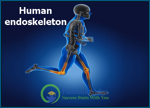
The human endoskeleton is the internal framework that provides support, protection, and mobility to the human body. It is made up of various components, primarily bones, joints, cartilage, and ligaments. This intricate system of structures plays a crucial role in maintaining the body’s shape, enabling movement, and safeguarding vital organs. Understanding the human endoskeleton is fundamental to appreciating the remarkable architecture that underpins human life.
Definition of Human Endoskeleton
The human endoskeleton is the internal skeleton that exists within the body, contrasting with exoskeletons found in some other organisms. It comprises bones, which are rigid structures made of a combination of organic collagen and inorganic minerals like calcium and phosphorus. These bones are interconnected through joints, held together by ligaments, and cushioned by cartilage.
Importance & Functions of the Endoskeleton
| Importance of Endoskeleton | Functions of Endoskeleton |
|---|---|
| Structural Support | Support the body, maintain posture, and enable an upright stance against gravity. |
| Protection of Organs | Shield delicate internal organs from injury and damage. |
| Facilitation of Movement | Work with muscles and joints to enable a wide range of movements and physical activities. |
| Muscle Attachment | Provide attachment points for muscles, allowing generation of force and movement. |
| Hematopoiesis | Some bones produce new blood cells through the process of hematopoiesis. |
| Mineral Storage | Act as reservoirs for essential minerals like calcium and phosphorus. |
| Mechanical Protection | Provide a protective buffer against external impacts and forces. |
| Growth and Development | Support growth and development during childhood and adolescence. |
| Shape and Aesthetic | Influence overall shape, physical characteristics, and appearance of an individual. |
| Adaptability and Evolution | Undergo adaptations over time, such as bipedalism, to influence human survival and success. |
| Bone Homeostasis | Continuously remodel to maintain bone density, strength, and adapt to changing demands. |
| Energy Storage | In certain circumstances, bones can serve as a storage site for energy in the form of fat. |
Composition of the Human Endoskeleton
The human endoskeleton is a complex system composed of four main components:
Bones
Bones are the rigid, hard structures that form the major part of the human endoskeleton. They are living tissues, constantly undergoing remodeling and regeneration. Bones vary in shape and size throughout the body and are classified into four types: long bones, short bones, flat bones, and irregular bones.
Joints
Joints are points of articulation where two or more bones meet. They allow for movement and flexibility in the skeleton. Joints can be classified into different types based on their structure and function, with the most common being synovial joints, cartilaginous joints, and fibrous joints.
Cartilage
Cartilage is a flexible and elastic connective tissue that covers the surface of bones at the joints. It acts as a shock absorber, reducing friction and preventing bones from rubbing against each other during movement.
Ligaments
Ligaments are tough bands of fibrous connective tissue that connect bones to other bones, providing stability and support to the joints. They play a crucial role in preventing excessive movement or dislocation of the bones.
Structure of Bones
Bones in the human endoskeleton come in various shapes and sizes, each with specific functions and characteristics:
Long Bones
Long bones are characterized by their elongated shape and consist of a shaft (diaphysis) and two ends (epiphyses). Examples of long bones include the femur, humerus, tibia, and fibula. They primarily function as levers for movement and are essential for weight-bearing activities.
Short Bones
Short bones are roughly cube-shaped and provide stability and support while facilitating limited gliding movements. Examples include the bones of the wrist (carpals) and ankle (tarsals).
Flat Bones
Flat bones are thin, flat, and usually curved. They offer protection to internal organs and provide a large surface area for muscle attachment. Examples include the skull bones (parietal and frontal), the scapula, and the sternum.
Irregular Bones
Irregular bones have complex shapes that don’t fit into the other categories. They serve a variety of functions, such as protection and muscle attachment. Examples include the vertebrae, facial bones (e.g., maxilla), and the hip bones (ilium, ischium, and pubis).
Bone Formation and Growth
The formation and growth of bones involve intricate processes that begin during fetal development and continue throughout life:
Ossification Process
The ossification process refers to the transformation of cartilage or fibrous tissue into bone. There are two primary types of ossification: endochondral ossification, which involves the replacement of cartilage, and intramembranous ossification, which occurs directly in connective tissue membranes.
Role of Osteoblasts and Osteoclasts
Osteoblasts are bone-forming cells responsible for synthesizing and depositing new bone tissue. On the other hand, osteoclasts are bone-resorbing cells that break down and remove old or damaged bone tissue. The balance between these two cell types is essential for maintaining bone health and density.
Factors Affecting Bone Growth
Several factors influence bone growth and development, such as genetics, nutrition, hormones, physical activity, and certain diseases. Proper nutrition, particularly an adequate intake of calcium, vitamin D, and other essential nutrients, is crucial for optimal bone health and growth.
Understanding the structure, formation, and functions of the human endoskeleton is fundamental for appreciating the intricacies of the human body and the role bones play in maintaining our physical abilities and overall well-being. With its remarkable combination of strength, flexibility, and adaptability, the human endoskeleton stands as a testament to the brilliance of nature’s design.
Major Bones of the Human Skeleton
The human skeleton is a marvel of engineering, providing the body with structure, support, and protection. It can be broadly divided into two main parts: the axial skeleton and the appendicular skeleton.
Axial Skeleton
The axial skeleton forms the central axis of the body and includes the following major bones:
- Skull:-
The skull is a complex structure consisting of 22 bones, including the cranium and facial bones. It encases and protects the brain and houses the sensory organs for vision, hearing, taste, and smell. - Spine (Vertebral Column):-
The spine, also known as the vertebral column, consists of 33 vertebrae stacked on top of one another. It provides support to the body and protects the spinal cord. The spine is divided into five regions: cervical, thoracic, lumbar, sacral, and coccygeal. - Ribs and Sternum:-
The ribcage comprises 12 pairs of ribs and the sternum (breastbone). The ribs protect the vital organs of the thoracic cavity, such as the heart and lungs, while the sternum serves as an anchor point for the ribs and several chest muscles.
Appendicular Skeleton
The appendicular skeleton includes the bones of the limbs and their associated girdles, which are the structures that connect the limbs to the axial skeleton.
- Upper Limb (Arm and Hand Bones):-
The upper limb consists of the humerus (upper arm bone), radius and ulna (forearm bones), carpals (wrist bones), metacarpals (palm bones), and phalanges (finger bones). It allows for a wide range of movements and precision grip. - Lower Limb (Leg and Foot Bones):-
The lower limb comprises the femur (thigh bone), tibia and fibula (leg bones), tarsals (ankle bones), metatarsals (sole bones), and phalanges (toe bones). The lower limb provides stability and supports body weight during standing and walking. - Shoulder Girdle (Clavicle and Scapula):-
The shoulder girdle consists of the clavicle (collarbone) and scapula (shoulder blade). It connects the upper limb to the axial skeleton and provides mobility to the arm. - Pelvic Girdle (Hip Bones):-
The pelvic girdle is composed of two hip bones, which fuse together to form the pelvis. It supports the weight of the upper body and protects the reproductive and digestive organs in the pelvic cavity.
Joint Types and Function
Joints are the points where two or more bones come together. They allow for movement and are classified into three main types based on their structure and function:
Synovial Joints
Synovial joints are the most common type of joint in the human body. They have a synovial cavity filled with synovial fluid that lubricates the joint, reducing friction during movement. Examples include the hinge joint in the elbow and the ball-and-socket joint in the hip.
Cartilaginous Joints
Cartilaginous joints have little to no movement and are connected by cartilage. There are two types: primary (synchondroses), found in growing bones, and secondary (symphyses), found in areas like the pubic symphysis.
Fibrous Joints
Fibrous joints are immovable and are connected by fibrous connective tissue. They provide stability and support to the bones. Examples include the sutures in the skull and the syndesmosis in the tibia and fibula.
Bone Development and Aging
Bone Remodeling
Bone remodeling is an ongoing process that involves the removal of old or damaged bone tissue by osteoclasts and the formation of new bone tissue by osteoblasts. This process maintains bone health and adapts the skeleton to mechanical stresses.
Bone Density and Aging
As we age, bone density tends to decrease, leading to a condition known as osteoporosis, where bones become fragile and prone to fractures. Adequate nutrition, exercise, and maintaining a healthy lifestyle can help mitigate bone density loss.
Common Bone-Related Disorders
Several bone-related disorders can affect the human skeleton, including osteoporosis, osteoarthritis, rheumatoid arthritis, and scoliosis. Early detection and appropriate treatment are essential in managing these conditions and maintaining bone health.
Understanding the major bones of the human skeleton, joint types, and bone development sheds light on the incredible complexity and functionality of the human body. From providing stability and protection to enabling intricate movements, the human skeletal system is a masterpiece of evolution and adaptation.
Role of Endoskeleton in Human Movement
The human endoskeleton plays a fundamental role in facilitating various movements and physical activities. It serves as a framework that provides support, stability, and protection to the body, allowing us to perform daily tasks and engage in a wide range of activities.
Support and Stability:
The endoskeleton’s primary function is to provide structural support to the body. The bones, in combination with ligaments and joints, create a stable framework that holds the body upright against the force of gravity. This support is essential for maintaining posture and balance, whether we are standing, walking, or sitting.
Muscle Attachment and Movement:
The endoskeleton acts as an anchor for muscles through tendons. When muscles contract, they exert force on the bones, causing movement at the joints. This muscle-bone interaction enables a vast array of movements, from simple actions like bending the fingers to complex movements like running, jumping, and dancing.
Protection of Internal Organs:
Another critical role of the endoskeleton is to protect vital internal organs from injury. For example, the skull safeguards the brain, the ribcage protects the heart and lungs, and the vertebral column shields the spinal cord. Without this protective function, the internal organs would be more susceptible to damage from external impacts.
Adaptations of the Human Skeleton
The human skeleton has undergone various adaptations over the course of evolution to meet the demands of the changing environment and lifestyle. Two notable adaptations include comparative anatomy with other animals and the development of bipedalism.
Comparative Anatomy with Other Animals
Comparative anatomy studies the similarities and differences in skeletal structures among different animal species. One of the most remarkable aspects of the human skeleton is its anatomical similarity to other primates, suggesting a shared evolutionary history. However, humans have distinct adaptations, such as a more upright posture, which differentiates them from other animals.
Bipedalism and Changes in Skeletal Structure
Bipedalism, the ability to walk on two feet, is one of the most significant adaptations in human evolution. As our ancestors transitioned from quadrupeds to bipeds, the human skeleton underwent several changes. These adaptations include modifications in the pelvic girdle, the curvature of the spine, and the positioning of the foramen magnum (the hole at the base of the skull where the spinal cord enters). These changes allowed for greater stability during upright walking and enabled the development of advanced cognitive and manipulative abilities.
Bone Health and Maintenance
Maintaining bone health is crucial for preventing injuries, promoting overall well-being, and preserving mobility throughout life. Several factors contribute to bone health, including nutrition, exercise, and lifestyle choices.
Importance of Nutrition
A balanced diet rich in essential nutrients is vital for maintaining healthy bones. Calcium, vitamin D, and vitamin K are particularly important for bone health. Calcium is a major component of bone tissue, while vitamin D aids in calcium absorption. Vitamin K helps regulate bone mineralization. Consuming dairy products, leafy greens, and fortified foods can help ensure adequate intake of these nutrients.
Exercise and Bone Density
Weight-bearing exercises, such as walking, jogging, dancing, and weightlifting, are beneficial for bone health. These activities stimulate bone remodeling and increase bone density, reducing the risk of osteoporosis and fractures. Additionally, resistance exercises that stress the bones, like strength training, contribute to bone health.
Common Bone Health Tips
- Avoid smoking and excessive alcohol consumption, as they can weaken bones.
- Practice good posture and body mechanics to reduce the risk of injury.
- Take precautions to prevent falls, especially in older adults, as falls can lead to fractures.
- Regularly visit a healthcare professional for bone health assessments and screenings.
Medical Imaging and Bone Examination
The human endoskeleton, hidden beneath the surface, holds many secrets about our physical health. Medical imaging techniques have revolutionized the way we examine and understand the human skeleton. Here are three commonly used imaging modalities for bone examination:
X-rays
X-rays are a widely used imaging tool for bone examination due to their ability to penetrate the body’s tissues, creating images of bones and other dense structures. X-rays are commonly employed to diagnose fractures, joint dislocations, and bone deformities. They provide a quick and relatively inexpensive way to visualize bones, making them a valuable tool in emergency medicine and orthopedics.
CT Scans (Computed Tomography)
CT scans, also known as CAT scans, use X-rays to create cross-sectional images of the body. CT scans offer a more detailed view of bones and can reveal fractures, tumors, infections, and complex bone injuries. The ability to visualize bone structures from different angles allows for better assessment of bone density and alignment.
MRI (Magnetic Resonance Imaging)
MRI utilizes powerful magnets and radio waves to generate detailed images of the internal structures of the body, including bones and soft tissues. While MRI is not the primary choice for examining bones, it excels at visualizing ligaments, tendons, and cartilage. It is particularly useful in diagnosing sports-related injuries and joint disorders.
Fun Facts about the Human Endoskeleton
The human endoskeleton is an incredible marvel of nature, serving as the backbone of our physical existence. Here are some fascinating facts about the human skeletal system:
Bone Strength and Composition
Despite being rigid and sturdy, bones are lightweight and strong. Their composition consists of organic collagen fibers, which provide flexibility, and inorganic minerals like calcium and phosphorus, which give bones their hardness. This unique combination of flexibility and strength allows bones to withstand considerable stress and forces.
Bone Regeneration Abilities
Bones possess the remarkable ability to regenerate and heal themselves. When a bone fractures, the body initiates a complex process of repair. Osteoblasts, bone-forming cells, deposit new bone tissue to bridge the gap between the fractured ends, eventually restoring the bone’s integrity. While bone healing takes time, the body’s regenerative capabilities ensure that most fractures can heal completely.
The Smallest Bone in the Human Body
The stapes, a tiny bone located in the middle ear, holds the distinction of being the smallest bone in the human body. This bone, resembling a stirrup, plays a critical role in transmitting sound vibrations from the eardrum to the inner ear.
Bones in the Hands and Feet
Approximately half of the bones in the human body are found in the hands and feet. Each hand contains 27 bones, while each foot houses 26 bones. The intricate structure of these bones allows for dexterity and precise movements, enabling us to perform a wide range of activities.
The Hyoid Bone
The hyoid bone is a unique bone that does not directly articulate with any other bones. Instead, it is suspended in the neck by ligaments and muscles. The hyoid bone serves as an anchor for the tongue and plays a crucial role in swallowing and speech production.
The human endoskeleton, an internal framework of bones, is a fascinating and intricate structure that enables our movements, protects our organs, and provides support to the body. Medical imaging techniques like X-rays, CT scans, and MRI have revolutionized our ability to examine and understand the skeleton, helping healthcare professionals diagnose and treat bone-related conditions effectively. Moreover, the endoskeleton’s strength, regenerative abilities, and unique bones make it a marvel of natural engineering, deserving of our awe and appreciation.