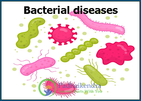
- Introduction to the Pectoral Girdle
- Definition and Overview
- Importance in the Skeletal System
- Anatomical Components
- Introduction to the Pectoral Girdle
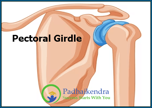
Welcome to the fascinating world of the pectoral girdle! In this article, we’ll embark on a journey to discover what this important part of our body is all about.
- Definition and Overview
So, what exactly is the pectoral girdle? Well, think of it as the upper part of your skeleton that’s like a bridge connecting your arms to your torso. It’s made up of bones and joints that play a vital role in your daily activities.
The pectoral girdle primarily consists of two main bones: the clavicle (collarbone) and the scapula (shoulder blade). These bones work together to support and move your arms, allowing you to do everything from giving a friendly wave to lifting heavy objects.
- Importance in the Skeletal System
Why is the pectoral girdle so important? Well, it’s like the cornerstone of your upper body. Just like the foundation of a building, your pectoral girdle provides support and stability to your upper limbs. Without it, your arms would feel like jelly, and you wouldn’t be able to perform many tasks we take for granted.
Imagine trying to reach for something on a high shelf or throwing a ball without your pectoral girdle. It would be quite challenging, right? That’s why understanding its structure and function is crucial to appreciating the complexity of the human body.
- Anatomical Components
Let’s break it down a bit further. Within the pectoral girdle, we have two main components: the clavicle and the scapula.
Clavicle (Collarbone): This slender bone runs horizontally across the front of your neck and chest, connecting your breastbone (sternum) to your shoulder. It acts as a strut, helping to hold your shoulder in place and allowing your arms to move freely. It’s like the bridge between your sternum and the rest of your arm.
Scapula (Shoulder Blade): The scapula is a flat, triangular bone located on your upper back, right behind your ribcage. It’s a bit like a wing, and it’s the main bone that moves when you raise or rotate your arm. It provides a stable base for the muscles that control your shoulder joint.
These two bones, along with the various joints and muscles connected to them, form the pectoral girdle. Together, they create a beautifully designed structure that enables you to perform countless activities in your daily life, from giving someone a hug to swinging a tennis racket.
Now that we’ve scratched the surface of the pectoral girdle, let’s dive deeper into its anatomy, functions, and the incredible ways it contributes to our daily lives and overall health.
- Anatomy of the Pectoral Girdle
- Clavicle (Collarbone)
- Structure and Characteristics
- Function
- Common Injuries and Disorders
- Anatomy of the Pectoral Girdle
Welcome back! Now, let’s zoom in on the pectoral girdle’s anatomy, starting with one of its key players: the clavicle, also known as the collarbone.
- Clavicle (Collarbone)
Structure and Characteristics
Picture a gentle, slender curve that runs right across the front of your neck and chest. That’s your clavicle, and it’s quite unique in its shape. The clavicle is typically an S-shaped bone that’s strong yet lightweight, making it a bit like nature’s own suspension bridge.
It’s not just any bone; the clavicle is the only long bone in the human body that lies horizontally. On one end, it attaches to your sternum (the breastbone), and on the other end, it meets your shoulder blade, forming a crucial connection between your trunk and your arm.
Function
Now, why is this bone so special? Well, it has a few essential jobs in the body. First and foremost, it acts as a sort of strut or support beam for your shoulder. It helps keep your shoulder in the right place and prevents it from sinking down. Imagine it like the beam that holds up a shelf.
Additionally, the clavicle plays a key role in the movement of your arm. It provides an anchor point for various muscles, allowing you to lift, swing, and rotate your arm. Without it, your shoulder wouldn’t be nearly as stable or mobile.
Common Injuries and Disorders
Like any part of your body, the clavicle isn’t invincible. It can face its fair share of challenges:
Fractures: Collarbone fractures are relatively common, especially in sports and accidents. These can happen if you fall on your shoulder or experience a direct blow. Thankfully, most collarbone fractures heal well with proper care, which might involve wearing a sling or, in some cases, surgery.
Dislocations: While not as common as fractures, dislocations of the clavicle can occur. This happens when the clavicle slips out of its normal position at the sternoclavicular joint or the acromioclavicular joint.
Arthritis: Just like other joints in your body, the sternoclavicular joint can develop arthritis over time, causing pain and limited movement.
Shoulder Impingement: Problems with the clavicle can also lead to shoulder impingement syndrome, where tendons in the shoulder get pinched and cause discomfort.
Understanding these potential issues can help you appreciate the importance of taking care of your collarbone and seeking prompt treatment if you ever face any of these problems.
So, there you have it—the clavicle, a unique and vital part of the pectoral girdle that keeps your shoulder in check and helps you move your arm gracefully. In our next section, we’ll shift our focus to another important component of the pectoral girdle: the scapula or shoulder blade. Stay tuned!
- Scapula (Shoulder Blade)
- Structure and Characteristics
- Function
- Common Injuries and Disorders
- Scapula (Shoulder Blade)
In our journey to understand the pectoral girdle, our spotlight now shines on the scapula, often known as the shoulder blade. This is another piece of the puzzle that makes our upper body work smoothly.
- Structure and Characteristics
Imagine a flat, triangular bone located on your upper back, right behind your ribcage—that’s the scapula. Its shape is like a big, angular wing, and it’s an interesting piece of the puzzle when it comes to your upper body’s design.
The scapula has a few standout features:
Spine: Running along its back, there’s a bony ridge called the spine of the scapula. This part not only helps the scapula keep its shape but also provides attachment points for important muscles.
Acromion Process: If you reach up and touch the tip of your shoulder, you’re actually feeling the acromion process of your scapula. It’s a bony projection that forms a part of your shoulder joint.
Glenoid Cavity: This is a shallow, socket-like depression on the side of the scapula. It’s a crucial piece of your shoulder joint, where your upper arm bone (the humerus) meets the scapula to form the glenohumeral joint. It’s like the hinge on a door, allowing your arm to swing in various directions.
- Function
Now, let’s talk about what this flat, wing-like bone does:
Stability and Movement: The scapula is like the foundation of your shoulder joint. It provides a stable base for the muscles that control your shoulder’s movements. When you raise your arm, rotate it, or even just move it a little bit, the scapula is involved. It’s like the stage on which the shoulder’s dance happens.
Muscle Attachment: Many important muscles attach to the scapula, including the trapezius, deltoid, and rotator cuff muscles. These muscles work together to give you the ability to move your arm in a wide range of motions, from reaching for a high shelf to throwing a ball.
- Common Injuries and Disorders
As with any part of your body, the scapula can face its share of challenges:
Fractures: While not as common as collarbone fractures, scapula fractures can happen due to accidents or trauma. They usually require medical attention to heal properly.
Scapular Dyskinesis: This is a condition where the normal movement of the scapula is altered. It can result from muscle imbalances or injuries and may lead to shoulder pain or dysfunction.
Shoulder Impingement Syndrome: Problems with the scapula’s movement can contribute to shoulder impingement, where the tendons in your shoulder get pinched. This can cause discomfort and limit your range of motion.
Understanding the structure, function, and potential issues with the scapula helps us appreciate how intricate our bodies are. It’s like a piece in a complex jigsaw puzzle, working in harmony with other bones and muscles to let us do everyday tasks and more.
As we continue our journey through the pectoral girdle, we’ll explore how the scapula and the clavicle work together and with other elements to make your upper body a well-oiled machine. Stay with us!
- Sternoclavicular Joint
- Structure and Location
- Function
- Movements and Range of Motion
- Sternoclavicular Joint
Now, let’s turn our attention to a joint that’s essential to the pectoral girdle’s functionality: the sternoclavicular joint. This joint might have a long name, but we’re going to break it down in simple terms.
- Structure and Location
The sternoclavicular joint is where the clavicle (collarbone) meets the sternum (breastbone). It’s like the point where two roads meet, except in this case, it’s where two bones come together.
Picture this: right in the middle of your chest, there’s a small, flat, and triangular bone called the sternum. Your sternum is important—it protects your heart and other vital organs. Now, think of the clavicle, which runs horizontally across your chest. At the front end of the clavicle, it joins the sternum at the sternoclavicular joint.
This joint is quite unique because it’s the only point where the clavicle connects to your main body frame, which is your sternum. It’s like the bridge that connects your shoulder to your chest.
- Function
So, what’s the job of the sternoclavicular joint? Well, it has a few important roles:
Stability: It helps hold your shoulder in place. Think of it as an anchor. Without this joint, your shoulder wouldn’t be firmly attached to your chest. It would be like trying to build a treehouse without nails.
Shock Absorption: It also has a bit of cushioning effect. When you lift something heavy or make sudden movements, the sternoclavicular joint absorbs some of the shock, protecting your shoulder and chest.
- Movements and Range of Motion
Now, let’s talk about what this joint allows you to do:
Elevation and Depression: You know when you raise your shoulders up and down? That’s thanks to the sternoclavicular joint. It allows your clavicle to move up and down, which is essential for tasks like shrugging your shoulders.
Protraction and Retraction: Ever push your shoulders forward or pull them back? That’s the joint at work again. It permits your clavicle to slide a bit forward and backward.
Rotation: This joint even helps with a bit of rotation. When you rotate your shoulder, the sternoclavicular joint allows your clavicle to pivot a bit.
Understanding the movements and range of motion of the sternoclavicular joint helps us appreciate its role in our everyday activities. Whether you’re reaching for something high on a shelf or simply adjusting your posture, this joint is quietly working behind the scenes.
In our exploration of the pectoral girdle, we’re piecing together how each part contributes to the overall function of your upper body. Next up, we’ll delve into the fascinating world of the acromioclavicular joint, another important player in this intricate system. Stay tuned for more!
III. Articulations of the Pectoral Girdle
- Acromioclavicular Joint
- Structure and Location
- Function
- Injuries and Treatment
III. Articulations of the Pectoral Girdle
Welcome back to our exploration of the pectoral girdle, the incredible framework that lets you move your upper body. In this section, we’re going to zoom in on one specific joint within the pectoral girdle: the acromioclavicular joint.
- Acromioclavicular Joint
Structure and Location
The acromioclavicular joint (AC joint, for short) is a small but crucial junction where two bones come together. These bones are the clavicle (collarbone) and the acromion process of the scapula (shoulder blade).
To visualize it, place your hand on your shoulder and feel that little bump at the tip of your shoulder blade. That bump is the acromion process. Now, follow your finger towards the front, and you’ll find the clavicle. Right where these two meet, you’ve discovered the AC joint. It’s like the intersection where two roads meet.
Function
What does the AC joint do? It plays a significant role in the stability and movement of your shoulder. Here’s how:
Stability: The AC joint helps keep your shoulder blade and collarbone connected, which is vital for shoulder stability. Without this connection, your shoulder wouldn’t be as steady, and everyday tasks like lifting and reaching would become tricky.
Shock Absorption: Just like the sternoclavicular joint, the AC joint also provides some shock absorption. When you lift something heavy or experience sudden jolts, it helps protect your shoulder from the impact.
Injuries and Treatment
Unfortunately, like many parts of the body, the AC joint can encounter problems:
Sprains: AC joint sprains are relatively common. They happen when there’s excessive force applied to the joint, like falling on your shoulder. Depending on the severity, these sprains can range from mild to severe.
Separation: AC joint separation is a more serious injury where the ligaments connecting the collarbone and shoulder blade are damaged. This can result in the collarbone moving out of its normal position, creating a noticeable bump on your shoulder.
Arthritis: Over time, the AC joint can develop arthritis, causing pain and limited mobility.
Treatment for AC joint injuries depends on their severity. Mild sprains might heal with rest and ice, while severe injuries may require bracing, physical therapy, or even surgery to restore normal function.
Understanding the structure, function, and potential issues with the AC joint helps us appreciate how this small but mighty joint plays a big role in our daily lives. It’s yet another part of the complex puzzle that is the pectoral girdle.
In our next segment, we’ll dive into the intriguing world of the glenohumeral joint, which is often called the shoulder joint. Stay tuned to uncover how this joint contributes to the incredible flexibility of your arm and shoulder movements!
- Glenohumeral Joint (Shoulder Joint)
- Structure and Components
- Function
- Range of Motion and Movements
- Common Shoulder Problems
- Glenohumeral Joint (Shoulder Joint)
Our journey through the pectoral girdle continues with a deep dive into the glenohumeral joint, often called the shoulder joint. Brace yourself for some fascinating insights into how this joint makes your arm a marvel of mobility.
- Structure and Components
The glenohumeral joint is a ball-and-socket joint, like the hinge on a door, but with way more range of motion. Let’s break down its structure:
Glenoid Cavity: The socket part of the joint is formed by the glenoid cavity, which is a shallow depression on the lateral edge of the scapula (shoulder blade). Think of it as a little dish.
Humerus: The ball part is the rounded head of the humerus, your upper arm bone. This head sits snugly in the glenoid cavity, like a golf ball fitting into a tee.
Now, imagine this setup. The glenoid cavity is shallow, but it’s surrounded by a rim of fibrous cartilage called the labrum, which adds depth and stability. This whole arrangement allows for a wide range of motion.
- Function
The glenohumeral joint is the star of the show when it comes to arm and shoulder movement. Its primary functions include:
Mobility: This joint is incredibly mobile, allowing your arm to move in all directions—up, down, forward, backward, and in circles. It’s like the universal joint in a car, enabling your arm to reach, lift, and rotate.
Stability: However, all that mobility comes with a trade-off. The glenohumeral joint is inherently less stable than some other joints, like the hip. To compensate, your body relies heavily on muscles, ligaments, and tendons to keep the joint in check.
- Range of Motion and Movements
The range of motion at the glenohumeral joint is astounding:
Flexion: This is when you raise your arm in front of you. Think of raising your hand to answer a question in class.
Extension: The opposite of flexion, this is when you move your arm backward, like reaching behind you.
Abduction: When you raise your arm out to the side, it’s abduction. Picture someone waving hello to you.
Adduction: The reverse of abduction, adduction is when you bring your arm back to your side.
Rotation: You can rotate your arm externally (turning your palm up) and internally (turning your palm down).
Circumduction: This is a fancy term for making a circular motion with your arm. Imagine stirring a giant pot of soup with your whole arm.
- Common Shoulder Problems
While the glenohumeral joint is impressive, it’s also prone to certain issues:
Rotator Cuff Tears: The muscles and tendons that stabilize the glenohumeral joint can tear or get injured, often due to overuse or trauma.
Shoulder Impingement Syndrome: This happens when the tendons in your shoulder get pinched as they pass through the narrow space beneath the acromion process.
Frozen Shoulder (Adhesive Capsulitis): Some people experience limited mobility and pain in their shoulder, which can be caused by inflammation and the thickening of the joint capsule.
Understanding the glenohumeral joint’s structure, function, and potential problems gives us insight into the incredible flexibility and occasional challenges of the shoulder. It’s like the master puppeteer behind your arm’s graceful movements.
In our next chapter, we’ll explore the costoclavicular joint, another crucial part of the pectoral girdle puzzle that adds stability to your shoulder. Stay tuned for more revelations!
- Costoclavicular Joint
- Structure and Function
- Costoclavicular Joint
Welcome to the next stop on our journey through the pectoral girdle: the costoclavicular joint. It might not be as famous as some other joints, but it plays a vital role in the stability of your shoulder.
- Structure and Function
The costoclavicular joint is a strong and sturdy joint found in your upper chest area. It’s where the clavicle (collarbone) meets the first rib and the cartilage that connects the first rib to your sternum (breastbone).
Now, let’s break down its structure:
Clavicle: We’ve met the clavicle before; it’s that curved bone running across the front of your neck and chest.
First Rib: The first rib is the shortest and sturdiest of all your ribs. It’s located right below the clavicle.
Cartilage: Between the first rib and the sternum, you’ll find cartilage that connects them, giving a bit of flexibility to this junction.
Now, what’s the job of this costoclavicular joint?
Stability: This joint is all about stability. It acts like an anchor point, keeping the clavicle firmly connected to your chest wall. Imagine it as the strong ropes holding a ship in place at the harbor.
Support: It provides crucial support for your shoulder girdle. When you lift heavy objects or perform activities that involve your arms, the costoclavicular joint helps distribute the load and prevents your shoulder from becoming wobbly.
In essence, the costoclavicular joint is like the unsung hero of the shoulder girdle, quietly doing its job to keep your upper body steady and strong.
While it may not have the same range of motion as other shoulder joints, it plays a crucial role in ensuring that your shoulder remains stable and functional. So, next time you lift something heavy or throw a ball, remember the role of the costoclavicular joint in making it all possible.
- Muscles and Ligaments of the Pectoral Girdle
- Muscles
- Deltoid Muscle
- Pectoral Muscles (Pectoralis Major and Minor)
- Trapezius Muscle
- Rotator Cuff Muscles
- Serratus Anterior Muscle
- Muscles and Ligaments of the Pectoral Girdle
Now, let’s dive into the muscular and ligamentous side of the pectoral girdle, where these mighty tissues work in harmony to help you perform a myriad of tasks. We’ll introduce you to some key players in this symphony of motion.
- Muscles
Deltoid Muscle
Meet the deltoid, a muscle that gives your shoulder its distinctive shape. It’s like the sleek, curved armor of a medieval knight. This muscle wraps around the shoulder joint, creating that beautifully rounded contour. The deltoid’s job is to lift your arm in various directions, making it essential for movements like lifting a weight or waving hello.
Pectoral Muscles (Pectoralis Major and Minor)
The pectoral muscles, often referred to as the pecs, are like two mighty wings that cover your chest. The pectoralis major is the larger and more superficial of the two, while the pectoralis minor lies beneath it. Together, they are responsible for various movements of the arm, such as flexion (bringing your arm forward) and adduction (bringing your arm closer to your body). They also contribute to the power of your shoulder and arm movements, like throwing a ball.
Trapezius Muscle
Imagine a trapeze artist gracefully swinging through the air. That’s the trapezius muscle in action, covering a large portion of your upper back and neck. It’s shaped like a trapezoid and plays a significant role in moving and stabilizing your shoulder blades. When you shrug your shoulders or pull them back, you’re thanking your trapezius for the effort.
Rotator Cuff Muscles
The rotator cuff muscles are like a team of tiny but crucial helpers. These four muscles—supraspinatus, infraspinatus, teres minor, and subscapularis—surround the shoulder joint, forming a cuff-like structure. They work together to stabilize and rotate the humerus (upper arm bone). These muscles are vital for the fine-tuned control of your shoulder’s movements, allowing you to throw a dart with precision or reach for a book on a high shelf without dislocating your shoulder.
Serratus Anterior Muscle
The serratus anterior, also known as the “boxer’s muscle,” is like a row of saw-like teeth along your ribs. It’s located beneath your scapula and plays a critical role in protraction (pushing your shoulder forward) and upward rotation of the scapula. This muscle helps your shoulder blade move smoothly against your ribcage, which is crucial for many arm movements, especially when you’re reaching out.
These muscles work together like a finely tuned orchestra, coordinating their efforts to create the beautiful symphony of movements that you perform every day. Without them, even simple actions like brushing your hair or lifting a cup of coffee would become challenging tasks.
In our exploration of the pectoral girdle, we’ve only just scratched the surface of the intricate web of muscles and ligaments that make it all work. In our next section, we’ll shift our focus to the ligaments of the pectoral girdle and understand their role in maintaining its stability. Stay tuned for more insights into the human body’s incredible design!
- Ligaments
- Coracoclavicular Ligaments
- Coracoacromial Ligament
- Glenohumeral Ligaments
- Muscles and Ligaments of the Pectoral Girdle
Welcome back to our journey through the pectoral girdle, where we’re now shining a spotlight on the crucial ligaments that help hold this intricate structure together. Ligaments are like the sturdy ropes that keep everything in place. Let’s meet some of these essential connectors:
- Ligaments
Coracoclavicular Ligaments
Imagine a bridge between your clavicle (collarbone) and another part of your shoulder. That’s precisely what the coracoclavicular ligaments are like. These ligaments connect the clavicle to a bony projection on the scapula called the coracoid process. These tough bands of tissue add strength and stability to the acromioclavicular joint, which is part of the shoulder girdle. They’re like the cables supporting a suspension bridge, ensuring your shoulder remains stable during various movements.
Coracoacromial Ligament
Picture a protective roof over your shoulder joint, shielding it from harm. The coracoacromial ligament is just that—a tough, protective structure that extends from the coracoid process to the acromion (a bony point of your scapula). It forms a sort of roof over the glenohumeral joint, helping prevent unwanted contact between your upper arm bone (humerus) and the acromion. Think of it as an umbrella, keeping your shoulder dry from potential injuries.
Glenohumeral Ligaments
These ligaments are the guardians of your shoulder’s front, middle, and back. They connect your humerus to the glenoid cavity of the scapula (shoulder blade), forming the glenohumeral joint. Think of them as strong, stretchy bands that hold your arm bone in place within the socket of the shoulder blade.
Anterior Glenohumeral Ligament: This one is at the front of your shoulder joint. It provides crucial stability, especially when you’re reaching forward or lifting something.
Middle Glenohumeral Ligament: As the name suggests, it’s in the middle. It helps with movements that involve abduction (raising your arm to the side) and external rotation (turning your arm outward).
Posterior Glenohumeral Ligament: Found at the back of the joint, this ligament comes into play when you internally rotate your arm (turning it inward). It’s like the safety net preventing over-rotation.
These ligaments work tirelessly, ensuring your shoulder joint remains stable and functional. Without them, the bones in your shoulder would be like puzzle pieces without glue, causing discomfort and limiting your range of motion.
So, whether you’re reaching for a book on a high shelf or throwing a ball with precision, remember the unheralded heroes—the ligaments of the pectoral girdle—making it all possible. In our next section, we’ll explore how these ligaments and muscles work together, creating harmony in your upper body’s movements. Stay tuned for more insights into the fascinating world of the human body!
- Function and Movement of the Pectoral Girdle
- Roles in Upper Limb Mobility
- Activities Involving the Pectoral Girdle
- Importance in Sports and Exercise
- Function and Movement of the Pectoral Girdle
In this section, we’re going to explore the dynamic aspects of the pectoral girdle—how it’s not just a static framework but a crucial player in your upper limb mobility, daily activities, and even sports and exercise. Let’s unravel the magic!
- Roles in Upper Limb Mobility
The pectoral girdle is like the anchor for your upper limbs, providing them with both support and mobility. Here’s how:
Shoulder Elevation: When you lift your arms to reach for the top shelf in your kitchen or wave hello, it’s the pectoral girdle that’s at work. Your scapula (shoulder blade) glides along the ribcage, giving your arm the freedom to rise.
Shoulder Depression: Conversely, when you lower your arms, the pectoral girdle ensures that your shoulder blades move downward smoothly. This action is essential for tasks like placing your hands on your hips or letting your arms hang by your sides.
Protraction and Retraction: Ever push something away from you or pull it closer? That’s the pectoral girdle’s job too. Protraction (pushing forward) and retraction (pulling backward) of your shoulder blades involve the scapula, which is a key component of the girdle.
- Activities Involving the Pectoral Girdle
Now, let’s talk about how you use your pectoral girdle in everyday life:
Getting Dressed: When you put on a shirt, blouse, or jacket, your pectoral girdle helps you move your arms to get those sleeves in place.
Driving: Turning the steering wheel, adjusting your side mirrors, and even signaling while driving—all involve your pectoral girdle’s coordinated efforts.
Cooking and Eating: Whether you’re chopping vegetables, lifting a pot, or bringing food to your mouth with a fork, your pectoral girdle is hard at work.
Playing Musical Instruments: If you’re a musician, your girdle plays a crucial role in strumming a guitar, playing the piano, or any other instrument requiring arm movement.
- Importance in Sports and Exercise
The pectoral girdle is a star player in the world of sports and exercise:
Swimming: Swimmers rely heavily on their pectoral girdles to perform various strokes, from freestyle to breaststroke. It’s what propels them through the water with power and grace.
Weightlifting: In sports like weightlifting and powerlifting, a strong pectoral girdle is essential for lifting heavy loads. It provides stability and support during exercises like bench presses and overhead lifts.
Yoga and Pilates: Even in activities focused on flexibility and balance, such as yoga and Pilates, the pectoral girdle plays a role in maintaining proper posture and achieving challenging poses.
Team Sports: In sports like basketball, volleyball, and baseball, your pectoral girdle comes into play when you throw, catch, or shoot. It’s like the hidden assistant making sure your arm movements are precise.
In essence, the pectoral girdle is your body’s versatile anchor. It enables you to perform a wide range of movements, from the simple acts of daily living to the complex actions required in sports and exercise. Understanding its function helps us appreciate the interconnectedness of our body’s systems, making it possible for us to enjoy an active and fulfilling life.
As we continue our exploration of the pectoral girdle, we’ll delve into its role in clinical considerations and its development and variations in the human body. So stay tuned for more insights into this incredible part of our anatomy!
- Clinical Considerations
- Common Pectoral Girdle Injuries
- Fractures and Dislocations
- Rotator Cuff Tears
- Shoulder Impingement Syndrome
- Clinical Considerations
In this chapter, we’re going to shift our focus to the real-world scenarios involving the pectoral girdle. While it’s a remarkable part of our anatomy, it’s not immune to injuries and issues. Let’s explore some common pectoral girdle injuries and conditions that can affect its function.
- Common Pectoral Girdle Injuries
Fractures and Dislocations
Clavicle Fractures: The clavicle (collarbone) is susceptible to fractures, often due to falls or direct blows. These fractures can be painful and may require immobilization with a sling or surgery in severe cases.
Shoulder Dislocations: Dislocations of the glenohumeral joint (shoulder joint) can occur, especially in high-impact sports or accidents. When the humerus (upper arm bone) pops out of its socket in the glenoid cavity, it’s both painful and unstable.
Rotator Cuff Tears
The rotator cuff muscles and tendons that stabilize the shoulder joint can become damaged over time or due to repetitive motion. Tears in these tissues can result in shoulder pain, weakness, and limited range of motion. Treatment options range from physical therapy to surgery, depending on the severity.
Shoulder Impingement Syndrome
In this condition, the tendons of the rotator cuff can become pinched or compressed under the acromion process of the scapula. This can lead to shoulder pain, especially when lifting your arm or reaching overhead. Treatment often involves rest, physical therapy, and sometimes corticosteroid injections.
Understanding these common injuries and conditions associated with the pectoral girdle is essential for maintaining shoulder health. Seeking prompt medical attention and following a tailored treatment plan can often lead to successful recovery.
It’s important to note that while these injuries can be painful and limiting, they are treatable, and many people recover fully with the right care and rehabilitation.
In our final section, we’ll take a closer look at the development and variations of the pectoral girdle in the human body, shedding light on how this structure can differ among individuals. Stay tuned for this fascinating exploration!
- Diagnostic Methods
- X-rays and Imaging
- Physical Examination
- Clinical Considerations
In this chapter, we’re going to shift our focus to the real-world scenarios involving the pectoral girdle. While it’s a remarkable part of our anatomy, it’s not immune to injuries and issues. Let’s explore some common pectoral girdle injuries and conditions that can affect its function.
- Diagnostic Methods
When it comes to identifying problems with the pectoral girdle, medical professionals have a few tricks up their sleeves. Let’s take a look at the diagnostic methods they use to get to the bottom of things:
X-rays and Imaging
Think of X-rays as your body’s own “photo shoot.” These high-energy waves pass through your body and create images of your bones on a special film or digital sensor. X-rays are particularly useful for spotting fractures, dislocations, and bone abnormalities in the pectoral girdle. It’s like shining a light in the dark to see what’s going on inside.
In addition to X-rays, doctors might use advanced imaging techniques like MRI (Magnetic Resonance Imaging) and CT (Computed Tomography) scans. These provide more detailed pictures of soft tissues, helping to diagnose issues like rotator cuff tears or ligament injuries. It’s like using a high-powered microscope to examine tiny details.
Physical Examination
Sometimes, a skilled doctor’s hands are the best diagnostic tool. They’ll use their fingers to feel for lumps, bumps, or abnormalities in the pectoral girdle area. They’ll also assess your range of motion, strength, and any pain or discomfort you might be experiencing. It’s like a detective examining a crime scene for clues.
During the physical examination, the doctor might perform specific tests like the “Neer Test” or the “Hawkins-Kennedy Test” to evaluate shoulder impingement or rotator cuff problems. These tests involve moving your arm in certain ways to elicit specific responses, helping the doctor pinpoint the issue.
Combining these diagnostic methods allows medical professionals to get a comprehensive view of the pectoral girdle’s health. This enables them to make accurate diagnoses and develop tailored treatment plans to help you get back to your normal activities.
Understanding these diagnostic methods empowers you to actively participate in your healthcare by discussing options with your healthcare provider and asking questions about your condition. In our final section, we’ll take a closer look at the development and variations of the pectoral girdle in the human body, shedding light on how this structure can differ among individuals. Stay tuned for this fascinating exploration!
- Treatment and Rehabilitation
- Conservative Approaches
- Surgical Interventions
- Physical Therapy
- Clinical Considerations
In the previous sections, we’ve explored the pectoral girdle’s structure, function, and potential issues. Now, let’s delve into how we can address these problems. Whether it’s through conservative methods, surgery, or physical therapy, there are ways to help your pectoral girdle get back in shape.
- Treatment and Rehabilitation
Conservative Approaches
When it comes to treating pectoral girdle injuries or conditions, conservative methods are often the first line of defense:
Rest and Immobilization: Sometimes, the best thing you can do for an injured pectoral girdle is to give it a break. Immobilizing the area with a sling or brace can promote healing, especially in cases of fractures or dislocations.
Medication: Pain relievers and anti-inflammatory drugs can help manage pain and reduce swelling associated with conditions like rotator cuff tears or shoulder impingement.
Physical Therapy: Physical therapists are like the architects of your recovery plan. They design customized exercises and stretches to improve strength, flexibility, and range of motion. These exercises target the muscles and structures of the pectoral girdle, gradually helping you regain function.
Heat and Cold Therapy: Applying heat or cold packs can provide relief from pain and inflammation. Heat can help relax tense muscles, while cold packs can reduce swelling.
Surgical Interventions
In some cases, conservative methods might not be enough, and surgical intervention becomes necessary:
Fracture Repair: Severe clavicle fractures or dislocated shoulders might require surgery to realign and stabilize the bones. Surgeons may use plates, screws, or pins to hold everything together.
Rotator Cuff Surgery: For significant rotator cuff tears, surgery may be recommended to repair the torn tendons. This can involve reattaching the tendon to the bone.
Joint Surgery: In cases of advanced arthritis or chronic instability, joint surgery, such as shoulder replacement or stabilization procedures, may be performed to restore function and alleviate pain.
Physical Therapy
Physical therapy isn’t just for recovery; it’s also an essential part of rehabilitation after surgical interventions. Your physical therapist will guide you through a structured program aimed at restoring strength, mobility, and function. They’ll also teach you how to perform movements correctly to prevent re-injury.
Therapy sessions can include a variety of exercises, such as range-of-motion exercises, strengthening exercises, and stretching routines. Your therapist will gradually increase the intensity as your pectoral girdle heals and becomes stronger.
Remember, everyone’s journey to recovery is unique, and the treatment plan will be tailored to your specific condition and needs. It’s essential to follow your healthcare provider’s advice, stick to your rehabilitation program, and be patient with the process.
In this final section, we’ll explore the fascinating world of the pectoral girdle’s development and variations in the human body. Understanding how this structure can differ among individuals can shed light on the diversity of our anatomy. Stay tuned for this enlightening exploration!
VII. Pectoral Girdle Development and Variations
- Embryonic Development
- Evolutionary Significance
- Anatomical Variations
VII. Pectoral Girdle Development and Variations
The pectoral girdle, as we’ve come to know it, is not just a static structure in the human body—it has a dynamic history of development and fascinating variations. In this section, we’ll uncover the story of how it forms in embryos, its evolutionary significance, and the interesting ways it can differ among individuals.
- Embryonic Development
The journey of the pectoral girdle begins during embryonic development. At a very early stage, the girdle begins to take shape. Here’s a simplified version of how it happens:
Mesenchyme: The pectoral girdle’s foundation starts with a type of embryonic tissue called mesenchyme. This tissue begins to condense and differentiate into the bones and connective tissues that will make up the girdle.
Clavicle and Scapula: The clavicle and scapula (collarbone and shoulder blade) start forming separately. The clavicle begins as a tiny cartilage model, which gradually ossifies (turns into bone) as the embryo develops. The scapula, on the other hand, starts as a flat plate of cartilage that goes through various transformations to become the familiar triangular shape we see in adults.
Muscles and Ligaments: Simultaneously, the muscles and ligaments that will attach to the pectoral girdle also begin their development. These include the muscles responsible for shoulder movement and stability.
The intricate dance of these developmental processes eventually gives rise to the fully formed pectoral girdle, ready to support the upper limbs and facilitate their impressive range of motion.
- Evolutionary Significance
The pectoral girdle’s evolution is a remarkable tale of adaptation. Over millions of years, it transformed from a simple structure in early vertebrates to the versatile and complex system we have today. Here’s a glimpse of its evolutionary journey:
Aquatic Beginnings: In early aquatic vertebrates, the pectoral girdle was primarily responsible for supporting the gills and stabilizing the body in water. It resembled a simple set of bony structures without the intricate mobility seen in land-dwelling animals.
Transition to Land: As vertebrates transitioned to life on land, the pectoral girdle had to adapt. It became more robust and gained greater mobility to support the weight of the body and facilitate activities like crawling and later, walking.
Flight and Swinging: In certain lineages, like birds and bats, the pectoral girdle underwent further modifications for flight. It became highly specialized, enabling the powered flapping of wings. In primates, including humans, the pectoral girdle adapted for swinging from tree branches, allowing for greater mobility in the trees.
The pectoral girdle’s evolutionary journey highlights the incredible adaptability of life forms over millions of years, shaping this structure into the versatile tool we now use for a wide range of activities.
- Anatomical Variations
In the world of anatomy, no two individuals are exactly alike. The pectoral girdle is no exception, and it can exhibit variations in several ways:
Clavicle Length: Some people have longer or shorter clavicles, which can affect the width of their shoulders and the range of motion in their arms.
Scapular Shape: Variations in the shape and orientation of the scapula can influence an individual’s posture and shoulder stability.
Muscle Attachment Points: The attachment points of muscles and ligaments on the pectoral girdle can differ among individuals, impacting their strength and mobility.
These variations, although subtle, contribute to the uniqueness of each person’s anatomy and can also play a role in how the pectoral girdle functions.
Understanding the development, evolution, and variations of the pectoral girdle adds depth to our appreciation of this remarkable anatomical structure. It reminds us that the human body is not just a static entity but a product of eons of evolution and adaptation. In our concluding section, we’ll summarize our journey through the pectoral girdle, highlighting its importance in our lives and the awe-inspiring complexity of our bodies. Stay with us for the grand finale!
Conclusion
Our exploration of the pectoral girdle, that often-overlooked but integral part of the human body, has taken us on a journey from its fundamental anatomy to its clinical considerations, developmental history, and intriguing variations. In conclusion, we’ve uncovered the following key takeaways:
Foundation of Mobility: The pectoral girdle is the cornerstone of upper limb mobility, allowing us to perform a myriad of tasks, from reaching for a book on a high shelf to throwing a ball with precision.
Clinical Realities: While a marvel of design, the pectoral girdle is not immune to injuries and conditions. Understanding diagnostic methods and treatment options empowers us to address and overcome these challenges effectively.
Development and Evolution: The embryonic development of the pectoral girdle is a testament to the remarkable process of growth and differentiation. Its evolution over millions of years showcases the adaptability of life forms and the diversity of anatomical structures.
Variations Among Us: Anatomical variations in the pectoral girdle remind us of the uniqueness of every individual’s body. Clavicle lengths, scapular shapes, and muscle attachments contribute to our distinct anatomical identities.
As we conclude our journey through the pectoral girdle, we are reminded of the awe-inspiring complexity of the human body. Each component, from the clavicle to the ligaments, muscles, and bones, plays a vital role in our ability to interact with the world around us.
May this exploration inspire curiosity and appreciation for the intricacies of our anatomy, and may it serve as a reminder that within every bone and joint lies a story of evolution, adaptation, and the wondrous capabilities of the human form.