- Introduction to Pulmonary Vein
- Definition and Overview
- Historical Significance
- Importance in Cardiovascular System
- Introduction to Pulmonary Vein
- Definition and Overview
The pulmonary vein, often called the “unsung hero” of the cardiovascular system, is a remarkable part of our bodies that plays a crucial role in keeping us alive. Let’s dive into the fascinating world of the pulmonary vein.
The pulmonary vein is not just one vein, but a group of veins that are responsible for a very important task – carrying oxygen-rich blood from our lungs back to our heart. This blood is loaded with life-giving oxygen, which our body’s cells need to function properly. Without the pulmonary vein, our bodies wouldn’t be able to get the oxygen they require to survive.
- Historical Significance
The history of the pulmonary vein is not as well-documented as some other parts of our anatomy, but its importance has been understood for centuries. Early anatomists and physicians knew that the heart and the blood vessels were vital to life, even if they didn’t fully comprehend the complexity of the pulmonary vein.
It wasn’t until the 16th century that Andreas Vesalius, a Belgian physician, made significant strides in understanding the structure of the heart and the blood vessels. His work laid the foundation for our modern understanding of the pulmonary vein and its role in the cardiovascular system.
- Importance in Cardiovascular System
Now, let’s talk about why the pulmonary vein is so important in our cardiovascular system. Our heart is like a powerful pump that constantly circulates blood throughout our body. But this blood isn’t just any fluid; it’s packed with nutrients and oxygen that our cells need to function correctly.
After blood circulates through our body and gives up its oxygen to our cells, it becomes oxygen-poor and filled with waste products like carbon dioxide. This “used” blood needs to be refreshed with oxygen so it can continue to nourish our cells. That’s where the pulmonary vein comes into play.
The pulmonary vein’s job is to collect this oxygen-poor blood from our lungs and bring it back to the heart. Once there, the heart pumps it into the lungs, where it picks up fresh oxygen and gets rid of the carbon dioxide. This revitalized blood is then sent back to the rest of the body, ready to support our cells and keep us healthy.
In summary, the pulmonary vein is a vital part of our cardiovascular system, ensuring that our body’s cells receive the oxygen they need to thrive. Its historical significance and its role in our bodies make it a fascinating subject to explore in our journey through the wonders of human anatomy.
- Anatomy of Pulmonary Vein
- Location and Position
- Structure and Layers
- Blood Flow and Circulation
- Anatomy of Pulmonary Vein
- Location and Position
Let’s take a closer look at where you can find the pulmonary vein in your body. This might sound like a GPS adventure, but it’s all about understanding your anatomy.
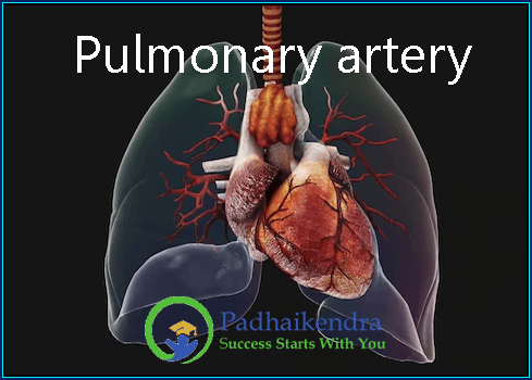
The pulmonary vein is like the gateway between your lungs and your heart. It’s located in your chest, snugly nestled within your lungs. There are usually four of them—one for each lung. These veins are like the couriers that bring freshly oxygenated blood back to your heart for a redeployment mission. They play a crucial role in the bloodstream’s grand adventure through your body.
- Structure and Layers
Now, let’s dissect the anatomy of the pulmonary vein—figuratively, of course. Imagine it as a tiny tube with a few layers. Each layer has a specific job to keep things running smoothly.
Innermost Layer (Endothelium): The innermost layer is super smooth and acts like a non-stick surface for blood cells. It helps prevent any unwanted stickiness in the vein and keeps the blood flowing freely.
Middle Layer (Smooth Muscle): Just like your biceps help you lift things, this layer contains smooth muscles that help regulate blood flow. They can squeeze and relax to control the amount of blood entering the vein.
Outer Layer (Connective Tissue): Think of this layer as the vein’s protective armor. It gives the vein strength and support, ensuring it doesn’t collapse or get damaged easily.
All these layers work together to ensure that your blood travels smoothly from your lungs back to your heart, maintaining the life-giving oxygen supply your body needs.
- Blood Flow and Circulation
The blood flow through the pulmonary vein is like a well-choreographed dance routine. Here’s how it goes:
Oxygenated blood from the tiny air sacs in your lungs (called alveoli) enters the pulmonary veins. This blood is as fresh as a spring morning.
The pulmonary veins collect this precious cargo and join together to form a larger vessel that leads straight to the left atrium of your heart. The left atrium is like the heart’s waiting room.
When your heart beats, the left atrium contracts, sending the oxygen-rich blood into the left ventricle, the heart’s main pumping chamber.
From there, the left ventricle contracts with a mighty push, sending the oxygenated blood out into your body through a major artery called the aorta. And off it goes to provide oxygen to every cell in your body.
So, you see, the pulmonary vein is like the lung’s personal FedEx, ensuring that the oxygen we breathe in gets where it needs to be. Understanding its anatomy helps us appreciate the intricate machinery that keeps us alive and well.
III. Function of Pulmonary Vein
- Transport of Oxygenated Blood
- Role in Pulmonary Circulation
- Connection to Left Atrium
III. Function of Pulmonary Vein
- Transport of Oxygenated Blood
Imagine your body as a bustling city with millions of tiny workers (cells) all needing a special ingredient to keep them going—oxygen. That’s where the pulmonary vein steps in as the oxygen delivery service.
Transporting Oxygen: The primary job of the pulmonary vein is to carry oxygen-rich blood from your lungs to your heart. Remember how you take a breath and your lungs fill up with oxygen? Well, that oxygen doesn’t just stay in your lungs; it needs to travel to every nook and cranny of your body. This is where the pulmonary vein plays its star role.
From Lungs to Heart: After your lungs have captured oxygen from the air, it’s dissolved into your blood. The pulmonary veins, which are like tiny highways, collect this freshly oxygenated blood and merge together into a larger vessel, heading towards your heart.
- Role in Pulmonary Circulation
Now, let’s take a closer look at what happens once the pulmonary vein delivers this oxygen-rich cargo to the heart.
Left Atrium’s Reception: The pulmonary vein ends its journey by pouring its load into the left atrium of your heart. Think of the left atrium as the heart’s waiting room. It’s where all the oxygenated blood gathers, patiently waiting for its turn to join the bloodstream highway.
The Beat Begins: When your heart does its rhythmic thumping, the left atrium contracts to send this oxygenated blood into the left ventricle, the heart’s main pumping chamber. This chamber is like a powerhouse, ready to send this precious cargo on its way.
- Connection to Left Atrium
The connection between the pulmonary vein and the left atrium is crucial. It’s like a secure gate that ensures the blood only goes where it’s supposed to.
One-Way Street: You see, there are valves in your heart that work like one-way doors. They allow blood to flow in one direction and prevent it from going backward. The connection between the pulmonary vein and the left atrium is guarded by one such valve, the mitral valve. This valve ensures that the oxygenated blood flows into the left atrium but can’t sneak back into the pulmonary vein.
In summary, the function of the pulmonary vein is a key player in your body’s oxygen delivery system. It carries the freshly oxygenated blood from your lungs, connects with the left atrium of your heart, and ensures that this vital cargo gets distributed to every cell, keeping you alive and thriving. Understanding its role in this intricate system helps us appreciate the amazing way our bodies work to keep us healthy.
- Pulmonary Vein Disorders and Diseases
- Pulmonary Vein Stenosis
- Causes and Risk Factors
- Symptoms and Diagnosis
- Treatment and Management
- Pulmonary Vein Disorders and Diseases
- Pulmonary Vein Stenosis
Pulmonary vein stenosis may sound like a mouthful, but it’s a condition worth understanding. It’s like a traffic jam in your body’s oxygen delivery system. Let’s break it down into causes, symptoms, and how to deal with it.
- Causes and Risk Factors
Traffic Jam in the Veins: Pulmonary vein stenosis happens when the pulmonary veins become too narrow or blocked. This can occur due to various reasons:
Congenital Defects: Sometimes, people are born with narrow pulmonary veins, which can lead to problems later in life.
Surgery or Procedures: Surgeries around the heart or the pulmonary veins themselves can sometimes cause scarring or narrowing.
Inflammation or Scarring: Conditions like inflammation or scarring can also constrict the veins.
- Symptoms and Diagnosis
Pulmonary vein stenosis doesn’t always announce itself loudly, but when it does, here’s what you might notice:
Breathing Troubles: Shortness of breath, especially during physical activity.
Fatigue: Feeling tired all the time, which can impact your daily life.
Chest Pain: Discomfort or pain in the chest, usually during exertion.
Diagnosis: Doctors use a combination of methods to diagnose pulmonary vein stenosis, including:
Imaging: Using techniques like echocardiography or CT scans to get a visual of the veins.
Catheterization: A tiny tube is threaded through the blood vessels to measure pressures and take samples.
Electrocardiogram (ECG or EKG): This records the heart’s electrical activity.
- Treatment and Management
Once diagnosed, there are a few ways to manage and treat pulmonary vein stenosis:
Medications: Doctors might prescribe medications to ease symptoms, reduce inflammation, or prevent clotting.
Angioplasty: In this procedure, a small balloon is inflated inside the narrowed vein to widen it.
Stent Placement: Sometimes, a stent (a mesh tube) is inserted to keep the vein open.
Surgery: In severe cases, surgery may be necessary to repair or replace the affected veins.
Ongoing Monitoring: Regular check-ups and imaging are essential to keep an eye on the condition’s progression.
Living with pulmonary vein stenosis can be challenging, but with the right medical care and lifestyle adjustments, many people can lead fulfilling lives. Remember, it’s vital to work closely with your healthcare team to determine the best treatment plan for your specific situation.
- Pulmonary Vein Thrombosis
- Causes and Risk Factors
- Symptoms and Diagnosis
- Treatment and Management
- Pulmonary Vein Disorders and Diseases
- Pulmonary Vein Thrombosis
Pulmonary vein thrombosis is like a traffic jam in a vital highway of your body, caused by clots blocking the road. Let’s delve into what causes this traffic jam, how to spot it, and what you can do about it.
- Causes and Risk Factors
Clots on the Highway: Pulmonary vein thrombosis occurs when blood clots, those tiny coagulated blobs, decide to set up camp in your pulmonary veins. Several factors can contribute to these clots forming:
Blood Clotting Disorders: Conditions that make your blood more prone to clotting.
Infections: Certain infections can trigger clot formation.
Injury or Surgery: Trauma, surgeries, or injuries can cause clots.
Catheter Use: Sometimes, medical procedures involving catheters can increase the risk.
- Symptoms and Diagnosis
Pulmonary vein thrombosis may not shout for attention, but it does leave some signs you should be aware of:
Breathing Trouble: Shortness of breath or difficulty in breathing.
Chest Pain: Pain in the chest, especially when you breathe deeply.
Coughing Up Blood: Sometimes, you may cough up blood, which is a definite red flag.
Fever: An unexplained fever can be a sign of an underlying issue.
Diagnosis: If you experience these symptoms, don’t wait—see a doctor. They’ll likely use these methods to diagnose pulmonary vein thrombosis:
Imaging: CT scans or MRIs can help visualize the clots and their location.
Blood Tests: These can reveal if there are any clotting disorders or infections.
- Treatment and Management
Managing pulmonary vein thrombosis is all about unclogging that traffic jam and ensuring smooth blood flow:
Anticoagulants: These are blood-thinning medications that help dissolve the clots and prevent new ones from forming.
Thrombolytics: In severe cases, doctors may use clot-busting medications to break down large clots quickly.
Pulmonary Angioplasty: If the clot is particularly stubborn, a procedure called pulmonary angioplasty can help. It involves inflating a balloon inside the vein to push the clot out of the way.
Surgery: Rarely, surgery may be necessary to remove large or life-threatening clots.
Prevention: Once treated, it’s essential to manage risk factors like blood clotting disorders and infections to prevent future clots.
Remember, pulmonary vein thrombosis is a serious condition that requires prompt medical attention. Early diagnosis and the right treatment plan can help clear the road and get your body’s traffic flowing smoothly again.
- Clinical Significance and Research
- Pulmonary Vein Ablation
- Imaging Techniques for Pulmonary Vein Assessment
- Ongoing Research and Advancements
- Clinical Significance and Research
- Pulmonary Vein Ablation
Let’s explore a high-tech procedure known as pulmonary vein ablation, discover how it can help, and peek into its significance in the world of cardiology.
What is Pulmonary Vein Ablation?
Pulmonary vein ablation, also known as pulmonary vein isolation (PVI), is a cutting-edge procedure used to treat a heart condition called atrial fibrillation (AFib). AFib is like a chaotic dance party in the heart’s upper chambers, causing irregular heartbeats. Pulmonary vein ablation aims to bring some order to this dance.
How Does It Work?
Imagine the pulmonary veins as the entrance gate to the heart’s upper chambers, known as the atria. In AFib, these veins can misbehave, sending erratic electrical signals to the heart. Pulmonary vein ablation steps in to calm things down.
During the procedure, a doctor carefully inserts catheters (thin tubes) into the heart through blood vessels. These catheters deliver either heat (radiofrequency ablation) or cold (cryoablation) to create scar tissue around the problematic veins. This scar tissue acts like a traffic cop, blocking the irregular electrical signals and restoring a regular heartbeat.
Significance in Cardiology:
Pulmonary vein ablation is a game-changer in the field of cardiology for several reasons:
Treating AFib: AFib can lead to serious complications like stroke, making effective treatment crucial.
Reducing Symptoms: Many patients experience symptoms like palpitations, fatigue, and shortness of breath due to AFib. Pulmonary vein ablation can significantly reduce or even eliminate these symptoms.
Improving Quality of Life: Restoring a regular heartbeat can improve a patient’s overall quality of life.
Less Reliance on Medications: Some patients may be able to reduce their reliance on medications after successful ablation.
- Imaging Techniques for Pulmonary Vein Assessment
When it comes to understanding the pulmonary veins, cutting-edge imaging techniques play a vital role. Let’s take a look at how these techniques are revolutionizing our ability to assess these crucial blood vessels.
- Echocardiography:
Echocardiography uses sound waves to create images of the heart and pulmonary veins. It’s a bit like sonar for your heart. This non-invasive method helps doctors see how blood flows through the pulmonary veins and can detect issues like blockages or clots.
- Cardiac MRI:
Cardiac magnetic resonance imaging (MRI) provides incredibly detailed images of the heart and pulmonary veins. It’s like taking a high-resolution photo of your heart’s inner workings. Cardiac MRI can help doctors diagnose pulmonary vein disorders and assess their severity.
- CT Angiography:
Computed tomography (CT) angiography involves taking a series of X-ray images to create a detailed map of the pulmonary veins. This method is especially useful for identifying structural issues or abnormalities in the pulmonary veins.
- Ongoing Research and Advancements
The world of pulmonary vein research is continually evolving, with scientists and doctors exploring new frontiers to improve our understanding and treatment of pulmonary vein-related conditions.
- Personalized Medicine:
Researchers are working to develop personalized treatment plans for individuals based on their unique pulmonary vein anatomy and conditions. This tailoring of care can lead to more effective treatments and better outcomes.
- Advanced Imaging:
Improvements in imaging technologies are making it easier for doctors to visualize and assess pulmonary veins with greater precision and accuracy.
- Minimally Invasive Procedures:
Ongoing research aims to make procedures like pulmonary vein ablation even less invasive, reducing patient discomfort and recovery times.
- Targeted Therapies:
Scientists are investigating targeted therapies that could help prevent conditions like thrombosis or stenosis from developing in the pulmonary veins.
In conclusion, clinical significance and research related to pulmonary veins are advancing our ability to diagnose and treat heart conditions. Techniques like pulmonary vein ablation and advanced imaging methods are improving the lives of patients with pulmonary vein disorders, and ongoing research promises even more exciting developments in the future.
- Surgical and Interventional Procedures
- Pulmonary Vein Isolation
- Pulmonary Vein Angioplasty
- Pulmonary Vein Transplant
- Surgical and Interventional Procedures
- Pulmonary Vein Isolation
Pulmonary vein isolation (PVI) is like the conductor of an orchestra, bringing harmony to a heart that’s out of rhythm. Let’s explore what PVI is, how it works, and when it’s used.
What is Pulmonary Vein Isolation?
Pulmonary vein isolation is a specialized procedure primarily used to treat a heart condition called atrial fibrillation (AFib). AFib is like a mischievous drummer in the heart’s rhythm band, causing irregular beats. PVI’s mission is to restore the heart’s natural tempo.
How Does It Work?
Imagine the pulmonary veins as the heart’s entry gates for oxygen-rich blood. In AFib, these veins send wonky electrical signals to the heart, leading to chaos. Pulmonary vein isolation steps in to bring order to the heart’s rhythm.
During PVI, a skilled cardiac electrophysiologist (heart rhythm specialist) uses catheters (thin tubes) to reach the heart. Once inside, they create scars around the pulmonary vein openings. These scars act like traffic barriers, preventing those pesky electrical signals from reaching the heart’s atria (upper chambers). As a result, the heart beats in a more regular and coordinated manner.
When is PVI Used?
Pulmonary vein isolation is typically considered when:
Medications to control AFib aren’t effective.
AFib symptoms significantly affect a patient’s quality of life.
There’s a high risk of stroke associated with AFib.
This procedure can help restore a regular heartbeat and improve the patient’s overall well-being.
- Pulmonary Vein Angioplasty
Pulmonary vein angioplasty is like a skilled road worker fixing a blocked highway. Let’s delve into what it is, why it’s needed, and how it’s done.
What is Pulmonary Vein Angioplasty?
Pulmonary vein angioplasty is a medical procedure used to treat blockages or narrowings in the pulmonary veins. These blockages can hinder the flow of oxygen-rich blood from the lungs to the heart, causing problems.
How Does It Work?
Imagine the pulmonary veins as the highways for oxygen-rich blood. Sometimes, these “vein highways” can get clogged with clots or narrowed due to scarring or other issues. Pulmonary vein angioplasty comes to the rescue.
During the procedure, a doctor inserts a catheter (a thin tube) into the blocked or narrowed vein. At the tip of the catheter, there’s a tiny balloon. When the catheter reaches the blockage, the balloon is inflated. This pushes the clot or plaque against the vein’s walls, opening it up and restoring the blood flow.
Why Is It Needed?
Pulmonary vein angioplasty is used when there are blockages or narrowings in the pulmonary veins that could affect oxygen delivery to the heart. It’s essential to keep these “vein highways” clear to maintain a healthy flow of oxygenated blood.
- Pulmonary Vein Transplant
Pulmonary vein transplant is a bit like transplanting a vital bridge to keep the traffic moving smoothly. Let’s explore what it entails, when it’s considered, and its significance.
What is Pulmonary Vein Transplant?
A pulmonary vein transplant is a highly specialized procedure where a damaged or non-functioning pulmonary vein is replaced with a healthy one from a donor. This is a relatively rare procedure, but it can be life-saving for those with severe pulmonary vein disorders.
When is Pulmonary Vein Transplant Considered?
Pulmonary vein transplants are typically considered in cases where:
The pulmonary vein is severely damaged or blocked beyond repair.
The patient’s life is at risk due to the condition of their pulmonary vein.
Other treatments, such as angioplasty or stent placement, are not viable options.
Significance of Pulmonary Vein Transplants:
Pulmonary vein transplants are significant because they can offer a second chance at life for individuals with severely damaged or non-functioning pulmonary veins. It’s a complex and intricate procedure that requires a highly skilled medical team and careful coordination with organ transplant organizations.
In conclusion, these surgical and interventional procedures, including pulmonary vein isolation, angioplasty, and transplantation, are essential tools in the medical toolkit for managing pulmonary vein disorders and ensuring the smooth flow of oxygenated blood in the cardiovascular system. They serve as lifelines for individuals facing these challenging conditions, helping them lead healthier and happier lives.
VII. Complications and Risks
- Complications of Pulmonary Vein Procedures
- Management of Complications
- Long-term Risks and Follow-up
VII. Complications and Risks
When it comes to medical procedures involving the pulmonary veins, it’s important to be aware of potential complications and long-term risks. Let’s explore what can go awry, how to manage these issues, and what to keep an eye on during follow-up.
- Complications of Pulmonary Vein Procedures
- Bleeding: During procedures like angioplasty or ablation, there’s a risk of bleeding at the catheter insertion site. While this is usually minor, it can be a concern.
- Blood Clots: Anytime you mess with blood vessels, there’s a small chance of blood clot formation, which can lead to more serious issues if not managed.
- Infection: Catheters entering the body can introduce bacteria, potentially causing infections.
- Damage to Surrounding Structures: In rare cases, procedures can inadvertently damage nearby structures, such as the heart’s electrical system.
- Procedure-Specific Complications: Each procedure comes with its own set of potential complications. For example, pulmonary vein isolation can sometimes result in complications like phrenic nerve injury, which affects breathing.
- Management of Complications
- Bleeding Control: If bleeding occurs, doctors can apply pressure to the insertion site to stop it. In severe cases, additional procedures may be needed to address the bleeding.
- Blood Thinners: To prevent and treat blood clots, doctors may prescribe blood-thinning medications.
- Antibiotics: If an infection is suspected, antibiotics can help clear it up.
- Repair Procedures: In the event of damage to surrounding structures, doctors may need to perform additional procedures to correct the issue.
- Immediate Attention: Any procedure-specific complications are addressed based on their nature. Doctors are trained to manage these situations promptly.
- Long-term Risks and Follow-up
Once the procedure is over, the journey isn’t complete. Long-term risks and follow-up care are essential components of a successful recovery.
- Recurrence: In some cases, pulmonary vein conditions can come back, requiring further treatment.
- Medication Management: Patients may need to continue taking medications to manage their condition, and it’s crucial to follow the prescribed regimen.
- Lifestyle Adjustments: Lifestyle changes, like maintaining a heart-healthy diet and regular exercise, can help reduce long-term risks.
- Follow-up Appointments: Regular check-ups with a healthcare provider are essential. These appointments allow doctors to monitor progress, make adjustments to treatment plans, and catch potential issues early.
- Risk Reduction: Patients should be aware of risk factors for pulmonary vein conditions and make efforts to reduce these risks, such as quitting smoking and managing conditions like high blood pressure or diabetes.
In summary, while procedures involving the pulmonary veins can be incredibly beneficial in treating various conditions, it’s crucial to acknowledge and address potential complications and long-term risks. With proper management and attentive follow-up care, individuals can navigate these challenges and maintain their cardiovascular health.
Conclusion
In conclusion, understanding the pulmonary vein and its intricate role in the cardiovascular system is essential for maintaining heart health. From its anatomy and vital functions to the procedures aimed at treating associated disorders, the pulmonary vein plays a pivotal role in ensuring our bodies receive the oxygen they need to thrive.
As we’ve explored, procedures like pulmonary vein isolation, angioplasty, and even transplants have revolutionized the field of cardiology, offering hope and improved quality of life for those dealing with pulmonary vein-related conditions.
However, it’s important to approach these medical interventions with awareness of potential complications and long-term risks. The diligent management of these challenges, coupled with regular follow-up care, is instrumental in achieving the best possible outcomes.
In the grand symphony of human health, the pulmonary vein is an unsung hero, quietly ensuring the continuous flow of oxygenated blood that sustains our lives. By staying informed, taking proactive steps to reduce risk factors, and collaborating closely with healthcare providers, we can navigate the complexities of pulmonary vein health and look forward to a healthier, happier future.